Calcium in PDB 5x7r: Crystal Structure of Paenibacillus Sp. 598K Alpha-1,6- Glucosyltransferase Complexed with Isomaltohexaose
Enzymatic activity of Crystal Structure of Paenibacillus Sp. 598K Alpha-1,6- Glucosyltransferase Complexed with Isomaltohexaose
All present enzymatic activity of Crystal Structure of Paenibacillus Sp. 598K Alpha-1,6- Glucosyltransferase Complexed with Isomaltohexaose:
3.2.1.20;
Protein crystallography data
The structure of Crystal Structure of Paenibacillus Sp. 598K Alpha-1,6- Glucosyltransferase Complexed with Isomaltohexaose, PDB code: 5x7r
was solved by
Z.Fujimoto,
N.Kishine,
N.Suzuki,
M.Momma,
H.Ichinose,
A.Kimura,
K.Funane,
with X-Ray Crystallography technique. A brief refinement statistics is given in the table below:
Resolution Low / High (Å)
|
152.46 /
1.95
|
Space group
|
C 2 2 21
|
Cell size a, b, c (Å), α, β, γ (°)
|
184.333,
271.260,
133.928,
90.00,
90.00,
90.00
|
R / Rfree (%)
|
17.8 /
20.8
|
Other elements in 5x7r:
The structure of Crystal Structure of Paenibacillus Sp. 598K Alpha-1,6- Glucosyltransferase Complexed with Isomaltohexaose also contains other interesting chemical elements:
Calcium Binding Sites:
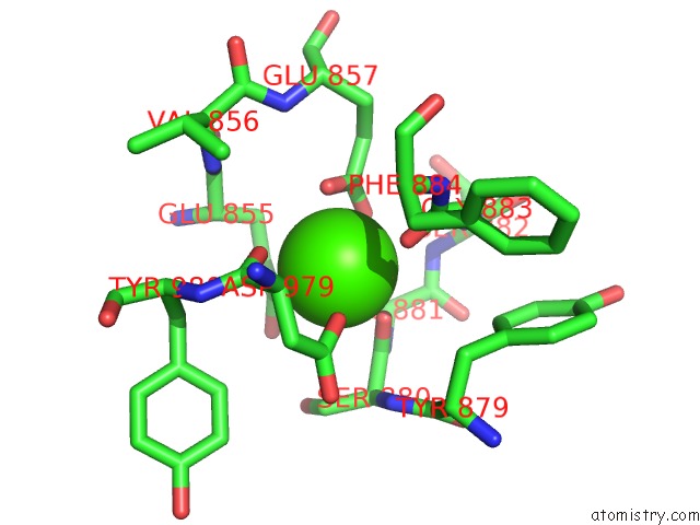
The binding sites of Calcium atom in the Crystal Structure of Paenibacillus Sp. 598K Alpha-1,6- Glucosyltransferase Complexed with Isomaltohexaose
(pdb code 5x7r). This binding sites where shown within
5.0 Angstroms radius around Calcium atom.
In total 6 binding sites of Calcium where determined in the
Crystal Structure of Paenibacillus Sp. 598K Alpha-1,6- Glucosyltransferase Complexed with Isomaltohexaose, PDB code: 5x7r:
Jump to Calcium binding site number:
1;
2;
3;
4;
5;
6;
Calcium binding site 1 out
of 6 in 5x7r
Go back to
Calcium Binding Sites List in 5x7r
Calcium binding site 1 out
of 6 in the Crystal Structure of Paenibacillus Sp. 598K Alpha-1,6- Glucosyltransferase Complexed with Isomaltohexaose
Mono view
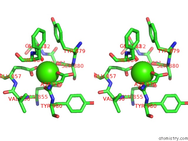
Stereo pair view
|
A full contact list of Calcium with other atoms in the Ca binding
site number 1 of Crystal Structure of Paenibacillus Sp. 598K Alpha-1,6- Glucosyltransferase Complexed with Isomaltohexaose within 5.0Å range:
probe
|
atom
|
residue
|
distance (Å)
|
B
|
Occ
|
A:Ca1301
b:33.7
occ:1.00
|
OE1
|
A:GLU855
|
2.2
|
33.1
|
1.0
|
OD1
|
A:ASP979
|
2.2
|
34.9
|
1.0
|
O
|
A:GLY883
|
2.4
|
34.3
|
1.0
|
O
|
A:SER880
|
2.4
|
36.9
|
1.0
|
OE1
|
A:GLU857
|
2.4
|
32.9
|
1.0
|
OE2
|
A:GLU857
|
2.6
|
33.9
|
1.0
|
O
|
A:ASP979
|
2.7
|
29.4
|
1.0
|
CD
|
A:GLU857
|
2.9
|
32.1
|
1.0
|
CD
|
A:GLU855
|
3.4
|
35.1
|
1.0
|
CG
|
A:ASP979
|
3.4
|
33.7
|
1.0
|
C
|
A:SER880
|
3.4
|
39.1
|
1.0
|
C
|
A:GLY883
|
3.5
|
34.2
|
1.0
|
C
|
A:ASP979
|
3.6
|
29.1
|
1.0
|
CA
|
A:ASP979
|
4.0
|
30.6
|
1.0
|
CB
|
A:GLU855
|
4.1
|
33.2
|
1.0
|
OE2
|
A:GLU855
|
4.2
|
36.5
|
1.0
|
OD2
|
A:ASP979
|
4.2
|
34.8
|
1.0
|
CG
|
A:GLU855
|
4.3
|
34.4
|
1.0
|
CA
|
A:SER880
|
4.3
|
38.3
|
1.0
|
N
|
A:GLY883
|
4.3
|
35.1
|
1.0
|
CB
|
A:ASP979
|
4.3
|
31.4
|
1.0
|
N
|
A:SER880
|
4.3
|
37.9
|
1.0
|
N
|
A:GLY881
|
4.3
|
42.6
|
1.0
|
CA
|
A:PHE884
|
4.3
|
34.2
|
1.0
|
N
|
A:PHE884
|
4.4
|
33.2
|
1.0
|
CG
|
A:GLU857
|
4.4
|
33.5
|
1.0
|
CB
|
A:SER880
|
4.4
|
38.4
|
1.0
|
CA
|
A:GLY881
|
4.5
|
41.6
|
1.0
|
CA
|
A:GLY883
|
4.5
|
35.5
|
1.0
|
CA
|
A:GLU855
|
4.5
|
31.8
|
1.0
|
N
|
A:TYR980
|
4.7
|
29.4
|
1.0
|
N
|
A:VAL856
|
4.8
|
31.4
|
1.0
|
N
|
A:SER882
|
4.8
|
39.1
|
1.0
|
CB
|
A:TYR879
|
4.8
|
38.4
|
1.0
|
C
|
A:GLY881
|
4.9
|
42.3
|
1.0
|
CB
|
A:PHE884
|
4.9
|
36.1
|
1.0
|
CB
|
A:TYR980
|
4.9
|
30.4
|
1.0
|
|
Calcium binding site 2 out
of 6 in 5x7r
Go back to
Calcium Binding Sites List in 5x7r
Calcium binding site 2 out
of 6 in the Crystal Structure of Paenibacillus Sp. 598K Alpha-1,6- Glucosyltransferase Complexed with Isomaltohexaose
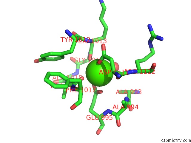
Mono view
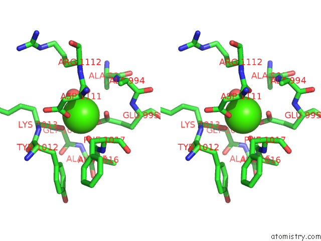
Stereo pair view
|
A full contact list of Calcium with other atoms in the Ca binding
site number 2 of Crystal Structure of Paenibacillus Sp. 598K Alpha-1,6- Glucosyltransferase Complexed with Isomaltohexaose within 5.0Å range:
probe
|
atom
|
residue
|
distance (Å)
|
B
|
Occ
|
A:Ca1302
b:28.8
occ:1.00
|
OD1
|
A:ASP1111
|
2.2
|
27.5
|
1.0
|
O
|
A:ALA1016
|
2.4
|
25.4
|
1.0
|
O
|
A:LYS1013
|
2.4
|
27.9
|
1.0
|
OE1
|
A:GLU995
|
2.4
|
23.7
|
1.0
|
O
|
A:HOH2713
|
2.5
|
24.8
|
1.0
|
OE2
|
A:GLU995
|
2.6
|
26.5
|
1.0
|
O
|
A:ASP1111
|
2.7
|
25.9
|
1.0
|
CD
|
A:GLU995
|
2.9
|
26.9
|
1.0
|
CG
|
A:ASP1111
|
3.3
|
30.1
|
1.0
|
C
|
A:LYS1013
|
3.5
|
29.3
|
1.0
|
C
|
A:ALA1016
|
3.6
|
24.4
|
1.0
|
C
|
A:ASP1111
|
3.6
|
26.4
|
1.0
|
CA
|
A:ASP1111
|
4.0
|
27.7
|
1.0
|
OD2
|
A:ASP1111
|
4.1
|
30.2
|
1.0
|
CB
|
A:ASP1111
|
4.3
|
28.4
|
1.0
|
N
|
A:LYS1013
|
4.3
|
28.0
|
1.0
|
CA
|
A:PHE1017
|
4.3
|
25.4
|
1.0
|
N
|
A:GLY1014
|
4.3
|
28.1
|
1.0
|
CA
|
A:LYS1013
|
4.4
|
29.4
|
1.0
|
N
|
A:PHE1017
|
4.4
|
25.3
|
1.0
|
CG
|
A:GLU995
|
4.4
|
25.8
|
1.0
|
N
|
A:ALA1016
|
4.4
|
27.2
|
1.0
|
CA
|
A:GLY1014
|
4.4
|
28.9
|
1.0
|
CA
|
A:ALA1016
|
4.6
|
26.6
|
1.0
|
CB
|
A:LYS1013
|
4.7
|
31.7
|
1.0
|
C
|
A:GLY1014
|
4.7
|
28.1
|
1.0
|
N
|
A:ALA994
|
4.8
|
25.6
|
1.0
|
CB
|
A:ALA993
|
4.8
|
27.2
|
1.0
|
CB
|
A:PHE1017
|
4.8
|
25.8
|
1.0
|
N
|
A:ARG1112
|
4.8
|
25.7
|
1.0
|
N
|
A:ALA1015
|
4.9
|
27.2
|
1.0
|
CB
|
A:TYR1012
|
4.9
|
26.3
|
1.0
|
|
Calcium binding site 3 out
of 6 in 5x7r
Go back to
Calcium Binding Sites List in 5x7r
Calcium binding site 3 out
of 6 in the Crystal Structure of Paenibacillus Sp. 598K Alpha-1,6- Glucosyltransferase Complexed with Isomaltohexaose
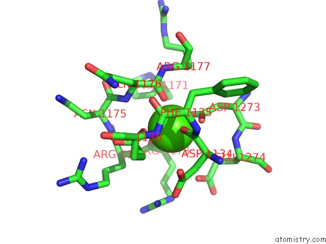
Mono view
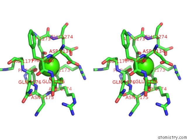
Stereo pair view
|
A full contact list of Calcium with other atoms in the Ca binding
site number 3 of Crystal Structure of Paenibacillus Sp. 598K Alpha-1,6- Glucosyltransferase Complexed with Isomaltohexaose within 5.0Å range:
probe
|
atom
|
residue
|
distance (Å)
|
B
|
Occ
|
A:Ca1303
b:33.5
occ:1.00
|
OE2
|
A:GLU1136
|
2.3
|
25.4
|
1.0
|
OD1
|
A:ASP1273
|
2.3
|
24.9
|
1.0
|
O
|
A:GLN1176
|
2.4
|
24.1
|
1.0
|
O
|
A:ASP1134
|
2.5
|
24.6
|
1.0
|
O
|
A:ARG1173
|
2.7
|
25.1
|
1.0
|
OD2
|
A:ASP1273
|
2.8
|
25.8
|
1.0
|
CG
|
A:ASP1273
|
2.9
|
25.4
|
1.0
|
CD
|
A:GLU1136
|
3.4
|
28.2
|
1.0
|
C
|
A:GLN1176
|
3.6
|
25.4
|
1.0
|
C
|
A:ASP1134
|
3.6
|
26.1
|
1.0
|
CG
|
A:GLU1136
|
3.8
|
27.3
|
1.0
|
C
|
A:ARG1173
|
3.9
|
24.1
|
1.0
|
CB
|
A:ASP1134
|
4.0
|
27.1
|
1.0
|
N
|
A:GLN1176
|
4.1
|
25.1
|
1.0
|
CA
|
A:ASP1134
|
4.2
|
26.3
|
1.0
|
CA
|
A:GLN1176
|
4.3
|
25.1
|
1.0
|
CB
|
A:PRO1171
|
4.4
|
29.1
|
1.0
|
CB
|
A:ASP1273
|
4.4
|
25.3
|
1.0
|
OE2
|
A:GLU1274
|
4.4
|
30.2
|
1.0
|
N
|
A:ARG1173
|
4.4
|
26.2
|
1.0
|
OE1
|
A:GLU1136
|
4.4
|
25.4
|
1.0
|
N
|
A:ARG1177
|
4.6
|
25.2
|
1.0
|
CA
|
A:ARG1173
|
4.6
|
26.0
|
1.0
|
N
|
A:PHE1135
|
4.7
|
25.1
|
1.0
|
CA
|
A:ARG1177
|
4.7
|
27.1
|
1.0
|
N
|
A:ASN1175
|
4.8
|
24.0
|
1.0
|
N
|
A:GLU1274
|
4.8
|
24.6
|
1.0
|
CB
|
A:ARG1174
|
4.8
|
26.0
|
1.0
|
CB
|
A:ARG1173
|
4.8
|
26.3
|
1.0
|
N
|
A:ARG1174
|
4.9
|
25.2
|
1.0
|
CG
|
A:ASP1134
|
4.9
|
27.4
|
1.0
|
N
|
A:GLU1136
|
4.9
|
24.9
|
1.0
|
CG
|
A:GLU1274
|
4.9
|
31.1
|
1.0
|
C
|
A:PHE1135
|
4.9
|
25.6
|
1.0
|
|
Calcium binding site 4 out
of 6 in 5x7r
Go back to
Calcium Binding Sites List in 5x7r
Calcium binding site 4 out
of 6 in the Crystal Structure of Paenibacillus Sp. 598K Alpha-1,6- Glucosyltransferase Complexed with Isomaltohexaose
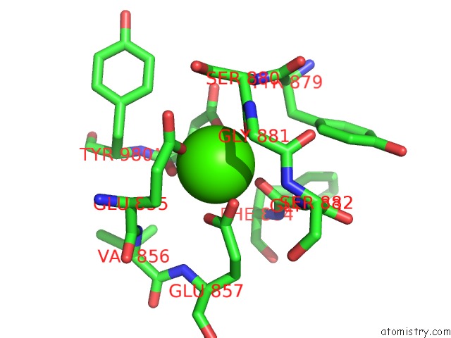
Mono view
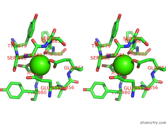
Stereo pair view
|
A full contact list of Calcium with other atoms in the Ca binding
site number 4 of Crystal Structure of Paenibacillus Sp. 598K Alpha-1,6- Glucosyltransferase Complexed with Isomaltohexaose within 5.0Å range:
probe
|
atom
|
residue
|
distance (Å)
|
B
|
Occ
|
B:Ca1301
b:30.3
occ:1.00
|
OE1
|
B:GLU855
|
2.2
|
30.3
|
1.0
|
OD1
|
B:ASP979
|
2.2
|
34.0
|
1.0
|
O
|
B:GLY883
|
2.3
|
28.7
|
1.0
|
O
|
B:SER880
|
2.5
|
32.6
|
1.0
|
OE1
|
B:GLU857
|
2.5
|
30.4
|
1.0
|
O
|
B:ASP979
|
2.5
|
26.3
|
1.0
|
OE2
|
B:GLU857
|
2.6
|
29.1
|
1.0
|
CD
|
B:GLU857
|
2.9
|
31.5
|
1.0
|
CD
|
B:GLU855
|
3.4
|
32.5
|
1.0
|
CG
|
B:ASP979
|
3.4
|
31.4
|
1.0
|
C
|
B:SER880
|
3.5
|
34.9
|
1.0
|
C
|
B:ASP979
|
3.5
|
27.4
|
1.0
|
C
|
B:GLY883
|
3.5
|
29.4
|
1.0
|
CA
|
B:ASP979
|
3.9
|
29.0
|
1.0
|
CB
|
B:GLU855
|
4.1
|
31.8
|
1.0
|
CB
|
B:ASP979
|
4.2
|
29.6
|
1.0
|
N
|
B:SER880
|
4.2
|
33.3
|
1.0
|
CA
|
B:SER880
|
4.2
|
34.2
|
1.0
|
OE2
|
B:GLU855
|
4.2
|
32.4
|
1.0
|
CG
|
B:GLU855
|
4.3
|
32.3
|
1.0
|
OD2
|
B:ASP979
|
4.3
|
30.7
|
1.0
|
N
|
B:GLY883
|
4.3
|
31.5
|
1.0
|
CB
|
B:SER880
|
4.3
|
33.0
|
1.0
|
N
|
B:GLY881
|
4.4
|
36.6
|
1.0
|
N
|
B:PHE884
|
4.4
|
27.7
|
1.0
|
CG
|
B:GLU857
|
4.4
|
30.6
|
1.0
|
CA
|
B:PHE884
|
4.4
|
28.2
|
1.0
|
CA
|
B:GLU855
|
4.5
|
31.2
|
1.0
|
CA
|
B:GLY881
|
4.5
|
37.4
|
1.0
|
CA
|
B:GLY883
|
4.5
|
28.3
|
1.0
|
N
|
B:TYR980
|
4.6
|
26.7
|
1.0
|
N
|
B:VAL856
|
4.7
|
29.1
|
1.0
|
CB
|
B:TYR980
|
4.7
|
29.4
|
1.0
|
CD2
|
B:TYR980
|
4.8
|
33.5
|
1.0
|
CB
|
B:TYR879
|
4.8
|
33.3
|
1.0
|
CB
|
B:PHE884
|
4.9
|
27.2
|
1.0
|
C
|
B:GLY881
|
4.9
|
37.6
|
1.0
|
C
|
B:GLU855
|
4.9
|
32.0
|
1.0
|
N
|
B:SER882
|
5.0
|
39.5
|
1.0
|
OG
|
B:SER880
|
5.0
|
33.2
|
1.0
|
|
Calcium binding site 5 out
of 6 in 5x7r
Go back to
Calcium Binding Sites List in 5x7r
Calcium binding site 5 out
of 6 in the Crystal Structure of Paenibacillus Sp. 598K Alpha-1,6- Glucosyltransferase Complexed with Isomaltohexaose
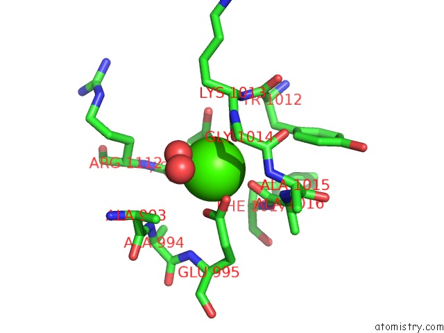
Mono view
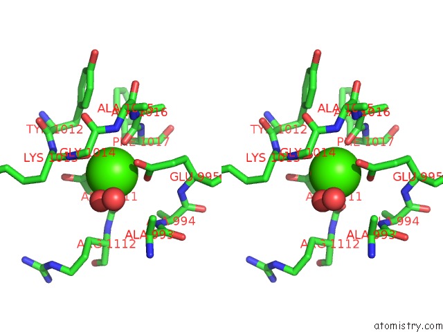
Stereo pair view
|
A full contact list of Calcium with other atoms in the Ca binding
site number 5 of Crystal Structure of Paenibacillus Sp. 598K Alpha-1,6- Glucosyltransferase Complexed with Isomaltohexaose within 5.0Å range:
probe
|
atom
|
residue
|
distance (Å)
|
B
|
Occ
|
B:Ca1302
b:30.4
occ:1.00
|
OD1
|
B:ASP1111
|
2.3
|
27.3
|
1.0
|
O
|
B:LYS1013
|
2.3
|
26.4
|
1.0
|
O
|
B:ALA1016
|
2.4
|
24.2
|
1.0
|
OE1
|
B:GLU995
|
2.4
|
23.7
|
1.0
|
O
|
B:HOH2734
|
2.5
|
25.2
|
1.0
|
OE2
|
B:GLU995
|
2.6
|
26.3
|
1.0
|
O
|
B:ASP1111
|
2.8
|
26.4
|
1.0
|
CD
|
B:GLU995
|
2.9
|
25.0
|
1.0
|
CG
|
B:ASP1111
|
3.4
|
29.2
|
1.0
|
C
|
B:LYS1013
|
3.4
|
28.3
|
1.0
|
C
|
B:ALA1016
|
3.6
|
23.8
|
1.0
|
C
|
B:ASP1111
|
3.7
|
25.9
|
1.0
|
CA
|
B:ASP1111
|
4.0
|
27.2
|
1.0
|
OD2
|
B:ASP1111
|
4.2
|
28.7
|
1.0
|
N
|
B:GLY1014
|
4.3
|
28.0
|
1.0
|
CB
|
B:ASP1111
|
4.3
|
27.6
|
1.0
|
N
|
B:ALA1016
|
4.3
|
27.3
|
1.0
|
N
|
B:LYS1013
|
4.3
|
25.5
|
1.0
|
CA
|
B:GLY1014
|
4.4
|
28.8
|
1.0
|
CA
|
B:PHE1017
|
4.4
|
24.5
|
1.0
|
CA
|
B:LYS1013
|
4.4
|
26.3
|
1.0
|
CG
|
B:GLU995
|
4.4
|
25.0
|
1.0
|
N
|
B:PHE1017
|
4.4
|
24.2
|
1.0
|
CA
|
B:ALA1016
|
4.6
|
26.1
|
1.0
|
O
|
B:HOH2781
|
4.7
|
43.4
|
1.0
|
C
|
B:GLY1014
|
4.7
|
28.9
|
1.0
|
N
|
B:ALA994
|
4.8
|
23.6
|
1.0
|
CB
|
B:ALA993
|
4.8
|
26.0
|
1.0
|
CB
|
B:LYS1013
|
4.8
|
27.4
|
1.0
|
N
|
B:ALA1015
|
4.8
|
27.3
|
1.0
|
N
|
B:ARG1112
|
4.9
|
25.7
|
1.0
|
CB
|
B:PHE1017
|
4.9
|
25.9
|
1.0
|
N
|
B:GLU995
|
5.0
|
21.9
|
1.0
|
CB
|
B:TYR1012
|
5.0
|
24.8
|
1.0
|
|
Calcium binding site 6 out
of 6 in 5x7r
Go back to
Calcium Binding Sites List in 5x7r
Calcium binding site 6 out
of 6 in the Crystal Structure of Paenibacillus Sp. 598K Alpha-1,6- Glucosyltransferase Complexed with Isomaltohexaose
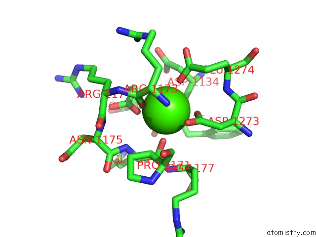
Mono view
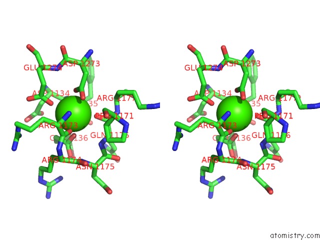
Stereo pair view
|
A full contact list of Calcium with other atoms in the Ca binding
site number 6 of Crystal Structure of Paenibacillus Sp. 598K Alpha-1,6- Glucosyltransferase Complexed with Isomaltohexaose within 5.0Å range:
probe
|
atom
|
residue
|
distance (Å)
|
B
|
Occ
|
B:Ca1303
b:33.5
occ:1.00
|
O
|
B:GLN1176
|
2.3
|
23.2
|
1.0
|
OD1
|
B:ASP1273
|
2.3
|
25.1
|
1.0
|
OE2
|
B:GLU1136
|
2.4
|
26.0
|
1.0
|
O
|
B:ASP1134
|
2.4
|
26.0
|
1.0
|
OE2
|
B:GLU1274
|
2.6
|
44.3
|
1.0
|
OD2
|
B:ASP1273
|
2.7
|
25.4
|
1.0
|
O
|
B:ARG1173
|
2.8
|
25.1
|
1.0
|
CG
|
B:ASP1273
|
2.9
|
25.1
|
1.0
|
CD
|
B:GLU1136
|
3.5
|
26.4
|
1.0
|
C
|
B:GLN1176
|
3.5
|
25.7
|
1.0
|
C
|
B:ASP1134
|
3.5
|
26.0
|
1.0
|
CG
|
B:GLU1136
|
3.9
|
26.3
|
1.0
|
CD
|
B:GLU1274
|
3.9
|
35.2
|
1.0
|
C
|
B:ARG1173
|
4.0
|
25.9
|
1.0
|
CB
|
B:ASP1134
|
4.0
|
27.1
|
1.0
|
N
|
B:GLN1176
|
4.1
|
25.9
|
1.0
|
CA
|
B:ASP1134
|
4.1
|
27.5
|
1.0
|
CA
|
B:GLN1176
|
4.4
|
25.3
|
1.0
|
CB
|
B:ASP1273
|
4.4
|
23.8
|
1.0
|
CB
|
B:PRO1171
|
4.4
|
30.7
|
1.0
|
N
|
B:ARG1173
|
4.5
|
27.8
|
1.0
|
N
|
B:ARG1177
|
4.5
|
26.9
|
1.0
|
OE1
|
B:GLU1136
|
4.6
|
24.2
|
1.0
|
CA
|
B:ARG1173
|
4.7
|
27.1
|
1.0
|
CA
|
B:ARG1177
|
4.7
|
27.3
|
1.0
|
N
|
B:PHE1135
|
4.7
|
23.1
|
1.0
|
OE1
|
B:GLU1274
|
4.7
|
34.9
|
1.0
|
N
|
B:GLU1274
|
4.7
|
25.5
|
1.0
|
CG
|
B:GLU1274
|
4.7
|
31.9
|
1.0
|
N
|
B:ASN1175
|
4.8
|
25.0
|
1.0
|
CB
|
B:ARG1174
|
4.8
|
24.2
|
1.0
|
CB
|
B:ARG1173
|
4.9
|
27.0
|
1.0
|
C
|
B:PHE1135
|
4.9
|
24.2
|
1.0
|
CG
|
B:ASP1134
|
4.9
|
29.7
|
1.0
|
CB
|
B:GLN1176
|
5.0
|
26.0
|
1.0
|
N
|
B:GLU1136
|
5.0
|
25.0
|
1.0
|
N
|
B:ARG1174
|
5.0
|
25.7
|
1.0
|
CB
|
B:ARG1177
|
5.0
|
28.0
|
1.0
|
|
Reference:
Z.Fujimoto,
N.Suzuki,
N.Kishine,
H.Ichinose,
M.Momma,
A.Kimura,
K.Funane.
Carbohydrate-Binding Architecture of the Multi-Modular Alpha-1,6-Glucosyltransferase From Paenibacillus Sp. 598K, Which Produces Alpha-1,6-Glucosyl-Alpha-Glucosaccharides From Starch Biochem. J. V. 474 2763 2017.
ISSN: ESSN 1470-8728
PubMed: 28698247
DOI: 10.1042/BCJ20170152
Page generated: Mon Jul 15 14:47:24 2024
|