Calcium »
PDB 5wzw-5xke »
5xag »
Calcium in PDB 5xag: Crystal Structure of Tubulin-Stathmin-Ttl-Compound Z2 Complex
Protein crystallography data
The structure of Crystal Structure of Tubulin-Stathmin-Ttl-Compound Z2 Complex, PDB code: 5xag
was solved by
H.Zhang,
C.Luo,
Y.Wang,
with X-Ray Crystallography technique. A brief refinement statistics is given in the table below:
| Resolution Low / High (Å) | 47.92 / 2.56 |
| Space group | P 21 21 21 |
| Cell size a, b, c (Å), α, β, γ (°) | 104.270, 156.117, 182.107, 90.00, 90.00, 90.00 |
| R / Rfree (%) | 21.1 / 25.5 |
Other elements in 5xag:
The structure of Crystal Structure of Tubulin-Stathmin-Ttl-Compound Z2 Complex also contains other interesting chemical elements:
| Magnesium | (Mg) | 5 atoms |
Calcium Binding Sites:
The binding sites of Calcium atom in the Crystal Structure of Tubulin-Stathmin-Ttl-Compound Z2 Complex
(pdb code 5xag). This binding sites where shown within
5.0 Angstroms radius around Calcium atom.
In total 3 binding sites of Calcium where determined in the Crystal Structure of Tubulin-Stathmin-Ttl-Compound Z2 Complex, PDB code: 5xag:
Jump to Calcium binding site number: 1; 2; 3;
In total 3 binding sites of Calcium where determined in the Crystal Structure of Tubulin-Stathmin-Ttl-Compound Z2 Complex, PDB code: 5xag:
Jump to Calcium binding site number: 1; 2; 3;
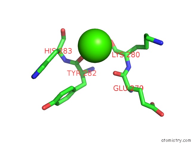
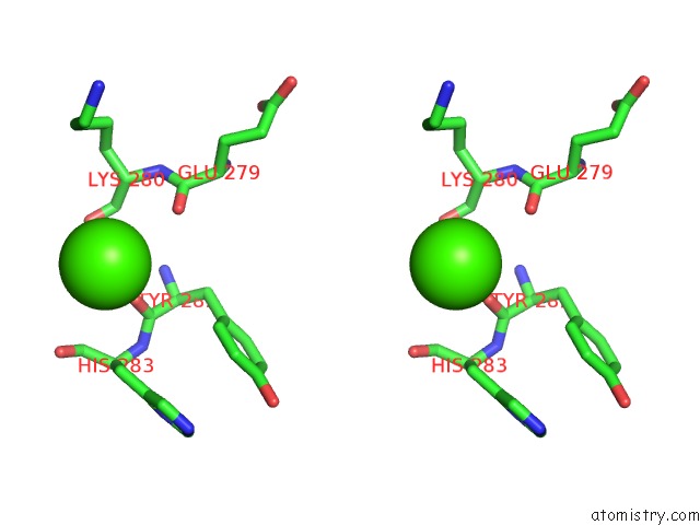
Calcium binding site 1 out of 3 in 5xag
Go back to
Calcium binding site 1 out
of 3 in the Crystal Structure of Tubulin-Stathmin-Ttl-Compound Z2 Complex
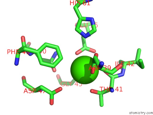
Mono view
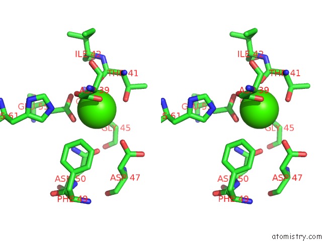
Stereo pair view
Mono view
Stereo pair view
A full contact list of Calcium with other atoms in the Ca binding
site number 1 of Crystal Structure of Tubulin-Stathmin-Ttl-Compound Z2 Complex within 5.0Å range:
|
Calcium binding site 2 out of 3 in 5xag
Go back to
Calcium binding site 2 out
of 3 in the Crystal Structure of Tubulin-Stathmin-Ttl-Compound Z2 Complex
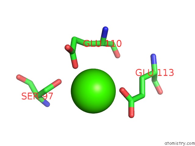
Mono view
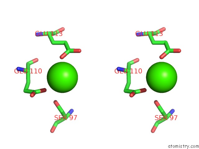
Stereo pair view
Mono view
Stereo pair view
A full contact list of Calcium with other atoms in the Ca binding
site number 2 of Crystal Structure of Tubulin-Stathmin-Ttl-Compound Z2 Complex within 5.0Å range:
|
Calcium binding site 3 out of 3 in 5xag
Go back to
Calcium binding site 3 out
of 3 in the Crystal Structure of Tubulin-Stathmin-Ttl-Compound Z2 Complex
Mono view
Stereo pair view
Mono view
Stereo pair view
A full contact list of Calcium with other atoms in the Ca binding
site number 3 of Crystal Structure of Tubulin-Stathmin-Ttl-Compound Z2 Complex within 5.0Å range:
|
Reference:
P.Zhou,
Y.Liang,
H.Zhang,
H.Jiang,
K.Feng,
P.Xu,
J.Wang,
X.Wang,
K.Ding,
C.Luo,
M.Liu,
Y.Wang.
Design, Synthesis, Biological Evaluation and Cocrystal Structures with Tubulin of Chiral Beta-Lactam Bridged Combretastatin A-4 Analogues As Potent Antitumor Agents Eur J Med Chem V. 144 817 2017.
ISSN: ISSN 1768-3254
PubMed: 29306206
DOI: 10.1016/J.EJMECH.2017.12.004
Page generated: Mon Jul 15 14:52:58 2024
ISSN: ISSN 1768-3254
PubMed: 29306206
DOI: 10.1016/J.EJMECH.2017.12.004
Last articles
Zn in 9MJ5Zn in 9HNW
Zn in 9G0L
Zn in 9FNE
Zn in 9DZN
Zn in 9E0I
Zn in 9D32
Zn in 9DAK
Zn in 8ZXC
Zn in 8ZUF