Calcium »
PDB 5wzw-5xke »
5xfm »
Calcium in PDB 5xfm: Crystal Structure of Beta-Arabinopyranosidase
Protein crystallography data
The structure of Crystal Structure of Beta-Arabinopyranosidase, PDB code: 5xfm
was solved by
K.Kato,
M.Okuyama,
M.Yao,
with X-Ray Crystallography technique. A brief refinement statistics is given in the table below:
| Resolution Low / High (Å) | 45.78 / 2.30 |
| Space group | P 1 |
| Cell size a, b, c (Å), α, β, γ (°) | 72.810, 90.510, 128.550, 105.50, 94.80, 96.15 |
| R / Rfree (%) | 20.7 / 25.3 |
Calcium Binding Sites:
The binding sites of Calcium atom in the Crystal Structure of Beta-Arabinopyranosidase
(pdb code 5xfm). This binding sites where shown within
5.0 Angstroms radius around Calcium atom.
In total 4 binding sites of Calcium where determined in the Crystal Structure of Beta-Arabinopyranosidase, PDB code: 5xfm:
Jump to Calcium binding site number: 1; 2; 3; 4;
In total 4 binding sites of Calcium where determined in the Crystal Structure of Beta-Arabinopyranosidase, PDB code: 5xfm:
Jump to Calcium binding site number: 1; 2; 3; 4;
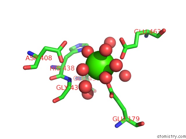
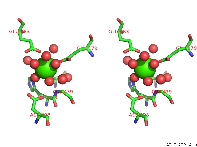
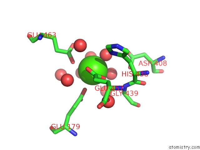
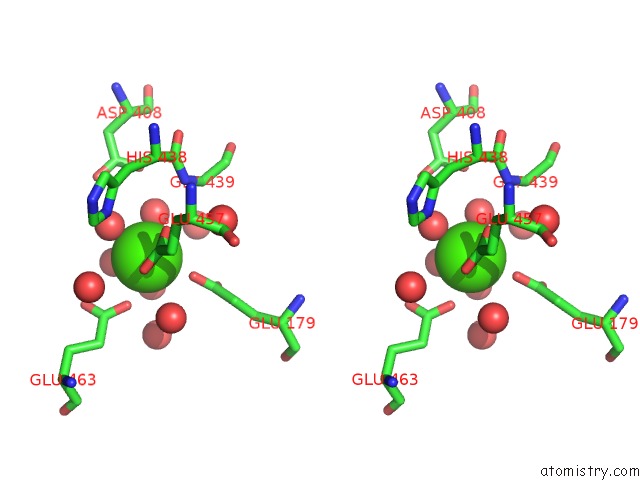
Calcium binding site 1 out of 4 in 5xfm
Go back to
Calcium binding site 1 out
of 4 in the Crystal Structure of Beta-Arabinopyranosidase
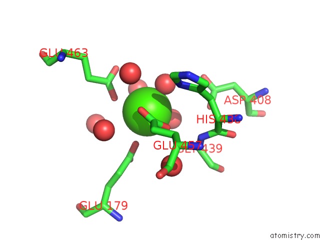
Mono view
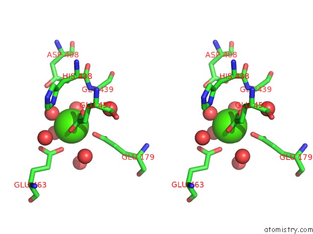
Stereo pair view
Mono view
Stereo pair view
A full contact list of Calcium with other atoms in the Ca binding
site number 1 of Crystal Structure of Beta-Arabinopyranosidase within 5.0Å range:
|
Calcium binding site 2 out of 4 in 5xfm
Go back to
Calcium binding site 2 out
of 4 in the Crystal Structure of Beta-Arabinopyranosidase
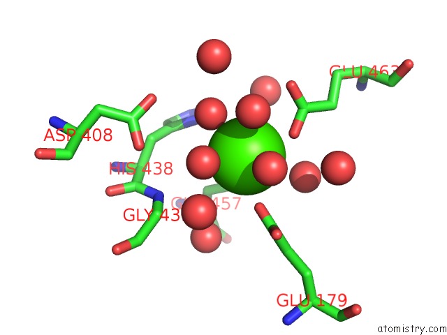
Mono view
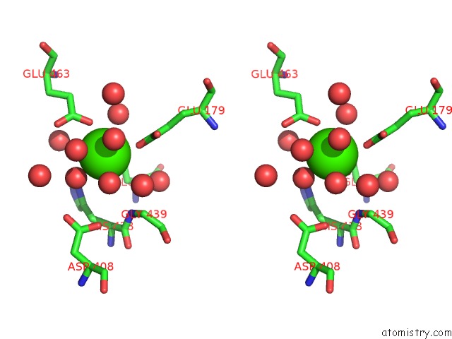
Stereo pair view
Mono view
Stereo pair view
A full contact list of Calcium with other atoms in the Ca binding
site number 2 of Crystal Structure of Beta-Arabinopyranosidase within 5.0Å range:
|
Calcium binding site 3 out of 4 in 5xfm
Go back to
Calcium binding site 3 out
of 4 in the Crystal Structure of Beta-Arabinopyranosidase
Mono view
Stereo pair view
Mono view
Stereo pair view
A full contact list of Calcium with other atoms in the Ca binding
site number 3 of Crystal Structure of Beta-Arabinopyranosidase within 5.0Å range:
|
Calcium binding site 4 out of 4 in 5xfm
Go back to
Calcium binding site 4 out
of 4 in the Crystal Structure of Beta-Arabinopyranosidase
Mono view
Stereo pair view
Mono view
Stereo pair view
A full contact list of Calcium with other atoms in the Ca binding
site number 4 of Crystal Structure of Beta-Arabinopyranosidase within 5.0Å range:
|
Reference:
A.Kikuchi,
M.Okuyama,
K.Kato,
S.Osaki,
M.Ma,
Y.Kumagai,
K.Matsunaga,
P.Klahan,
T.Tagami,
M.Yao,
A.Kimura.
A Novel Glycoside Hydrolase Family 97 Enzyme: Bifunctional Beta-L-Arabinopyranosidase/ Alpha-Galactosidase From Bacteroides Thetaiotaomicron. Biochimie V. 142 41 2017.
ISSN: ISSN 1638-6183
PubMed: 28804002
DOI: 10.1016/J.BIOCHI.2017.08.003
Page generated: Mon Jul 15 14:55:38 2024
ISSN: ISSN 1638-6183
PubMed: 28804002
DOI: 10.1016/J.BIOCHI.2017.08.003
Last articles
Zn in 9MJ5Zn in 9HNW
Zn in 9G0L
Zn in 9FNE
Zn in 9DZN
Zn in 9E0I
Zn in 9D32
Zn in 9DAK
Zn in 8ZXC
Zn in 8ZUF