Calcium »
PDB 5xkf-5xu9 »
5xs8 »
Calcium in PDB 5xs8: Crystal Structure of Solute-Binding Protein Complexed with Unsaturated Chondroitin Disaccharide with Two Sulfate Groups at C-4 and C-6 Positions of Galnac
Protein crystallography data
The structure of Crystal Structure of Solute-Binding Protein Complexed with Unsaturated Chondroitin Disaccharide with Two Sulfate Groups at C-4 and C-6 Positions of Galnac, PDB code: 5xs8
was solved by
S.Oiki,
R.Kamochi,
B.Mikami,
K.Murata,
W.Hashimoto,
with X-Ray Crystallography technique. A brief refinement statistics is given in the table below:
| Resolution Low / High (Å) | 40.40 / 1.95 |
| Space group | P 1 |
| Cell size a, b, c (Å), α, β, γ (°) | 49.741, 69.208, 165.967, 89.99, 90.04, 90.03 |
| R / Rfree (%) | 17.3 / 21.6 |
Calcium Binding Sites:
The binding sites of Calcium atom in the Crystal Structure of Solute-Binding Protein Complexed with Unsaturated Chondroitin Disaccharide with Two Sulfate Groups at C-4 and C-6 Positions of Galnac
(pdb code 5xs8). This binding sites where shown within
5.0 Angstroms radius around Calcium atom.
In total 4 binding sites of Calcium where determined in the Crystal Structure of Solute-Binding Protein Complexed with Unsaturated Chondroitin Disaccharide with Two Sulfate Groups at C-4 and C-6 Positions of Galnac, PDB code: 5xs8:
Jump to Calcium binding site number: 1; 2; 3; 4;
In total 4 binding sites of Calcium where determined in the Crystal Structure of Solute-Binding Protein Complexed with Unsaturated Chondroitin Disaccharide with Two Sulfate Groups at C-4 and C-6 Positions of Galnac, PDB code: 5xs8:
Jump to Calcium binding site number: 1; 2; 3; 4;
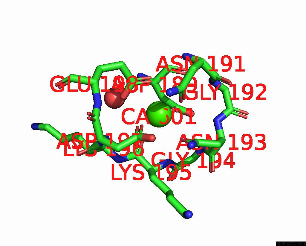
Calcium binding site 1 out of 4 in 5xs8
Go back to
Calcium binding site 1 out
of 4 in the Crystal Structure of Solute-Binding Protein Complexed with Unsaturated Chondroitin Disaccharide with Two Sulfate Groups at C-4 and C-6 Positions of Galnac
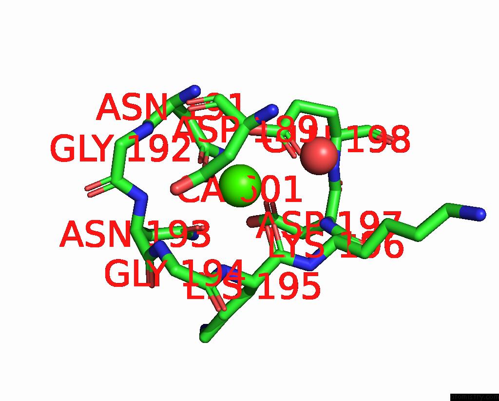
Mono view
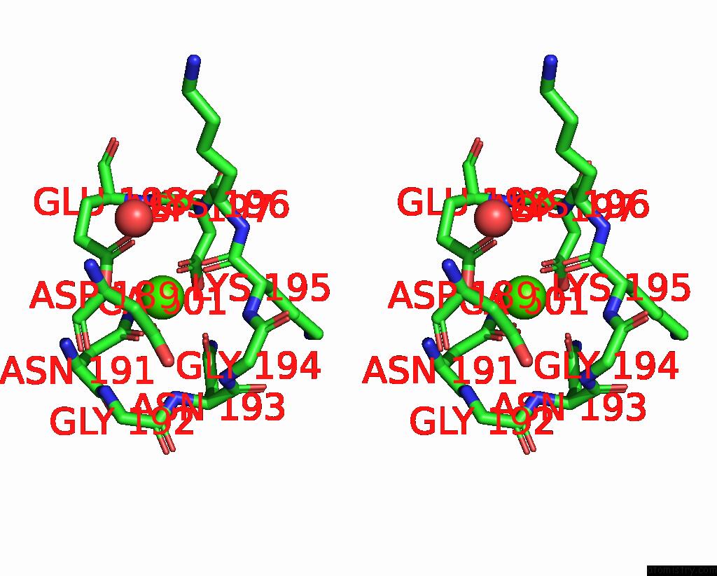
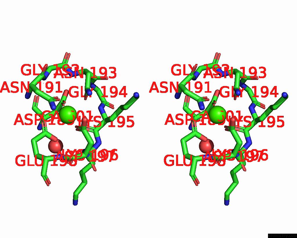
Stereo pair view
Mono view
Stereo pair view
A full contact list of Calcium with other atoms in the Ca binding
site number 1 of Crystal Structure of Solute-Binding Protein Complexed with Unsaturated Chondroitin Disaccharide with Two Sulfate Groups at C-4 and C-6 Positions of Galnac within 5.0Å range:
|
Calcium binding site 2 out of 4 in 5xs8
Go back to
Calcium binding site 2 out
of 4 in the Crystal Structure of Solute-Binding Protein Complexed with Unsaturated Chondroitin Disaccharide with Two Sulfate Groups at C-4 and C-6 Positions of Galnac
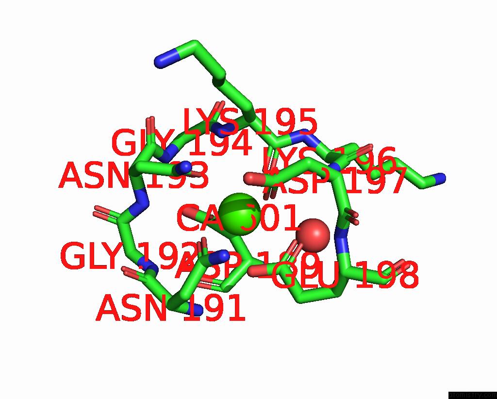
Mono view
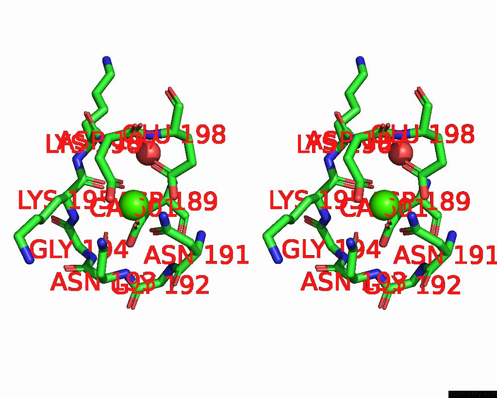
Stereo pair view
Mono view
Stereo pair view
A full contact list of Calcium with other atoms in the Ca binding
site number 2 of Crystal Structure of Solute-Binding Protein Complexed with Unsaturated Chondroitin Disaccharide with Two Sulfate Groups at C-4 and C-6 Positions of Galnac within 5.0Å range:
|
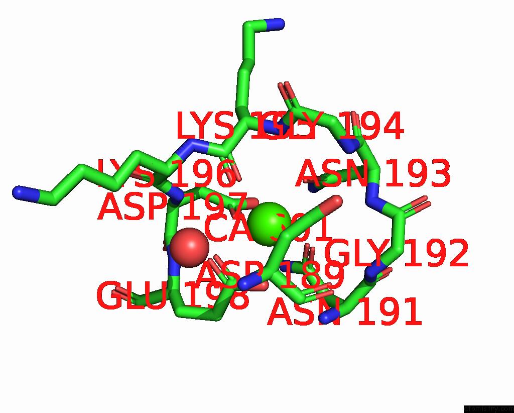
Calcium binding site 3 out of 4 in 5xs8
Go back to
Calcium binding site 3 out
of 4 in the Crystal Structure of Solute-Binding Protein Complexed with Unsaturated Chondroitin Disaccharide with Two Sulfate Groups at C-4 and C-6 Positions of Galnac
Mono view
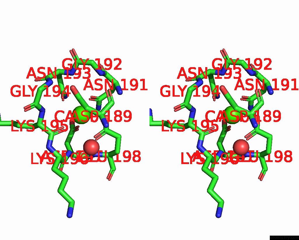
Stereo pair view
Mono view
Stereo pair view
A full contact list of Calcium with other atoms in the Ca binding
site number 3 of Crystal Structure of Solute-Binding Protein Complexed with Unsaturated Chondroitin Disaccharide with Two Sulfate Groups at C-4 and C-6 Positions of Galnac within 5.0Å range:
|
Calcium binding site 4 out of 4 in 5xs8
Go back to
Calcium binding site 4 out
of 4 in the Crystal Structure of Solute-Binding Protein Complexed with Unsaturated Chondroitin Disaccharide with Two Sulfate Groups at C-4 and C-6 Positions of Galnac
Mono view
Stereo pair view
Mono view
Stereo pair view
A full contact list of Calcium with other atoms in the Ca binding
site number 4 of Crystal Structure of Solute-Binding Protein Complexed with Unsaturated Chondroitin Disaccharide with Two Sulfate Groups at C-4 and C-6 Positions of Galnac within 5.0Å range:
|
Reference:
S.Oiki,
R.Kamochi,
B.Mikami,
K.Murata,
W.Hashimoto.
Alternative Substrate-Bound Conformation of Bacterial Solute-Binding Protein Involved in the Import of Mammalian Host Glycosaminoglycans. Sci Rep V. 7 17005 2017.
ISSN: ESSN 2045-2322
PubMed: 29208901
DOI: 10.1038/S41598-017-16801-8
Page generated: Wed Jul 9 11:46:47 2025
ISSN: ESSN 2045-2322
PubMed: 29208901
DOI: 10.1038/S41598-017-16801-8
Last articles
Cl in 5L5WCl in 5L5X
Cl in 5L5V
Cl in 5L5U
Cl in 5L5S
Cl in 5L5T
Cl in 5L5R
Cl in 5L5Q
Cl in 5L5P
Cl in 5L5O