Calcium »
PDB 5xuf-5yj7 »
5xwa »
Calcium in PDB 5xwa: Crystal Structure of Porcine Pancreatic Trypsin with Tripeptide Inhibitor, Pry, at pH 10
Enzymatic activity of Crystal Structure of Porcine Pancreatic Trypsin with Tripeptide Inhibitor, Pry, at pH 10
All present enzymatic activity of Crystal Structure of Porcine Pancreatic Trypsin with Tripeptide Inhibitor, Pry, at pH 10:
3.4.21.4;
3.4.21.4;
Protein crystallography data
The structure of Crystal Structure of Porcine Pancreatic Trypsin with Tripeptide Inhibitor, Pry, at pH 10, PDB code: 5xwa
was solved by
N.S.Saikhedkar,
A.S.Bhoite,
A.P.Giri,
K.A.Kulkarni,
with X-Ray Crystallography technique. A brief refinement statistics is given in the table below:
| Resolution Low / High (Å) | 41.58 / 1.90 |
| Space group | P 61 2 2 |
| Cell size a, b, c (Å), α, β, γ (°) | 83.165, 83.165, 135.232, 90.00, 90.00, 120.00 |
| R / Rfree (%) | 15.3 / 18.2 |
Calcium Binding Sites:
The binding sites of Calcium atom in the Crystal Structure of Porcine Pancreatic Trypsin with Tripeptide Inhibitor, Pry, at pH 10
(pdb code 5xwa). This binding sites where shown within
5.0 Angstroms radius around Calcium atom.
In total only one binding site of Calcium was determined in the Crystal Structure of Porcine Pancreatic Trypsin with Tripeptide Inhibitor, Pry, at pH 10, PDB code: 5xwa:
In total only one binding site of Calcium was determined in the Crystal Structure of Porcine Pancreatic Trypsin with Tripeptide Inhibitor, Pry, at pH 10, PDB code: 5xwa:
Calcium binding site 1 out of 1 in 5xwa
Go back to
Calcium binding site 1 out
of 1 in the Crystal Structure of Porcine Pancreatic Trypsin with Tripeptide Inhibitor, Pry, at pH 10
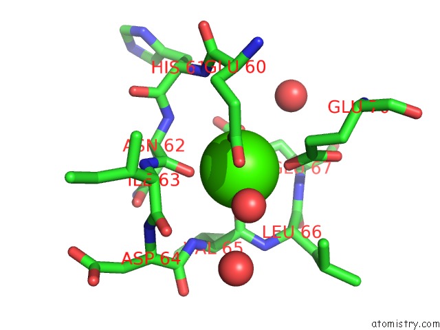
Mono view
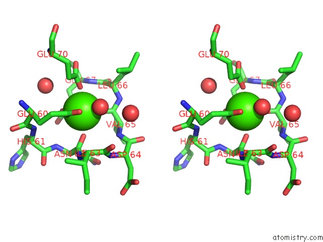
Stereo pair view
Mono view
Stereo pair view
A full contact list of Calcium with other atoms in the Ca binding
site number 1 of Crystal Structure of Porcine Pancreatic Trypsin with Tripeptide Inhibitor, Pry, at pH 10 within 5.0Å range:
|
Reference:
N.S.Saikhedkar,
R.S.Joshi,
A.S.Bhoite,
R.Mohandasan,
A.K.Yadav,
M.Fernandes,
K.A.Kulkarni,
A.P.Giri.
Tripeptides Derived From Reactive Centre Loop of Potato Type II Protease Inhibitors Preferentially Inhibit Midgut Proteases of Helicoverpa Armigera. Insect Biochem. Mol. Biol. V. 95 17 2018.
ISSN: ISSN 1879-0240
PubMed: 29486250
DOI: 10.1016/J.IBMB.2018.02.001
Page generated: Mon Jul 15 15:11:02 2024
ISSN: ISSN 1879-0240
PubMed: 29486250
DOI: 10.1016/J.IBMB.2018.02.001
Last articles
Zn in 9J0NZn in 9J0O
Zn in 9J0P
Zn in 9FJX
Zn in 9EKB
Zn in 9C0F
Zn in 9CAH
Zn in 9CH0
Zn in 9CH3
Zn in 9CH1