Calcium »
PDB 5xuf-5yj7 »
5yf1 »
Calcium in PDB 5yf1: Crystal Structure of CARNMT1 Bound to Carnosine and Sfg
Enzymatic activity of Crystal Structure of CARNMT1 Bound to Carnosine and Sfg
All present enzymatic activity of Crystal Structure of CARNMT1 Bound to Carnosine and Sfg:
2.1.1.22;
2.1.1.22;
Protein crystallography data
The structure of Crystal Structure of CARNMT1 Bound to Carnosine and Sfg, PDB code: 5yf1
was solved by
R.Cao,
H.Li,
with X-Ray Crystallography technique. A brief refinement statistics is given in the table below:
| Resolution Low / High (Å) | 41.59 / 2.40 |
| Space group | P 61 2 2 |
| Cell size a, b, c (Å), α, β, γ (°) | 128.125, 128.125, 324.643, 90.00, 90.00, 120.00 |
| R / Rfree (%) | 16.8 / 20.1 |
Calcium Binding Sites:
The binding sites of Calcium atom in the Crystal Structure of CARNMT1 Bound to Carnosine and Sfg
(pdb code 5yf1). This binding sites where shown within
5.0 Angstroms radius around Calcium atom.
In total 2 binding sites of Calcium where determined in the Crystal Structure of CARNMT1 Bound to Carnosine and Sfg, PDB code: 5yf1:
Jump to Calcium binding site number: 1; 2;
In total 2 binding sites of Calcium where determined in the Crystal Structure of CARNMT1 Bound to Carnosine and Sfg, PDB code: 5yf1:
Jump to Calcium binding site number: 1; 2;
Calcium binding site 1 out of 2 in 5yf1
Go back to
Calcium binding site 1 out
of 2 in the Crystal Structure of CARNMT1 Bound to Carnosine and Sfg
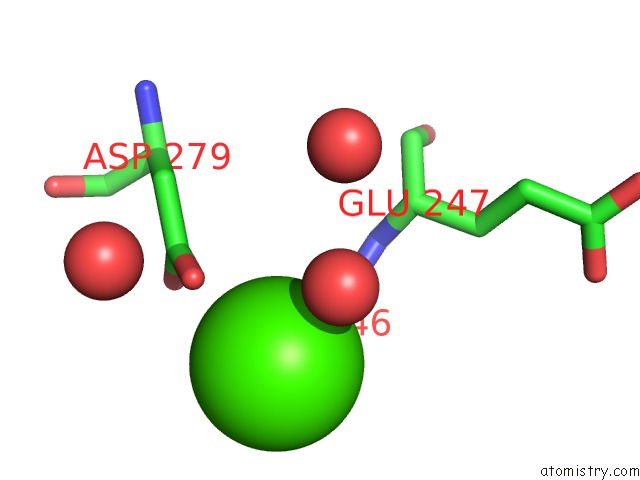
Mono view
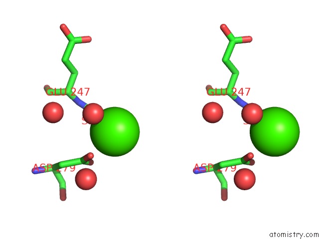
Stereo pair view
Mono view
Stereo pair view
A full contact list of Calcium with other atoms in the Ca binding
site number 1 of Crystal Structure of CARNMT1 Bound to Carnosine and Sfg within 5.0Å range:
|
Calcium binding site 2 out of 2 in 5yf1
Go back to
Calcium binding site 2 out
of 2 in the Crystal Structure of CARNMT1 Bound to Carnosine and Sfg
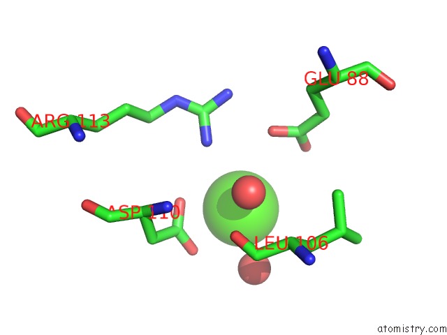
Mono view
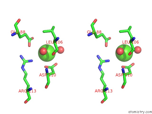
Stereo pair view
Mono view
Stereo pair view
A full contact list of Calcium with other atoms in the Ca binding
site number 2 of Crystal Structure of CARNMT1 Bound to Carnosine and Sfg within 5.0Å range:
|
Reference:
R.Cao,
X.Zhang,
X.Liu,
Y.Li,
H.Li.
Molecular Basis For Histidine N1 Position-Specific Methylation By CARNMT1. Cell Res. V. 28 494 2018.
ISSN: ISSN 1748-7838
PubMed: 29463897
DOI: 10.1038/S41422-018-0003-0
Page generated: Mon Jul 15 15:20:45 2024
ISSN: ISSN 1748-7838
PubMed: 29463897
DOI: 10.1038/S41422-018-0003-0
Last articles
Zn in 9MJ5Zn in 9HNW
Zn in 9G0L
Zn in 9FNE
Zn in 9DZN
Zn in 9E0I
Zn in 9D32
Zn in 9DAK
Zn in 8ZXC
Zn in 8ZUF