Calcium »
PDB 5yjd-5z4p »
5yjd »
Calcium in PDB 5yjd: Structural Insights Into the Crispr-Cas-Associated Ribonuclease Activity of Staphylococcus Epidermidis CSM3
Protein crystallography data
The structure of Structural Insights Into the Crispr-Cas-Associated Ribonuclease Activity of Staphylococcus Epidermidis CSM3, PDB code: 5yjd
was solved by
Y.Q.Zhao,
Y.J.Gu,
X.F.Zhu,
W.Cheng,
with X-Ray Crystallography technique. A brief refinement statistics is given in the table below:
| Resolution Low / High (Å) | 52.88 / 2.26 |
| Space group | I 21 21 21 |
| Cell size a, b, c (Å), α, β, γ (°) | 58.623, 105.760, 155.977, 90.00, 90.00, 90.00 |
| R / Rfree (%) | 19.7 / 24.1 |
Calcium Binding Sites:
The binding sites of Calcium atom in the Structural Insights Into the Crispr-Cas-Associated Ribonuclease Activity of Staphylococcus Epidermidis CSM3
(pdb code 5yjd). This binding sites where shown within
5.0 Angstroms radius around Calcium atom.
In total only one binding site of Calcium was determined in the Structural Insights Into the Crispr-Cas-Associated Ribonuclease Activity of Staphylococcus Epidermidis CSM3, PDB code: 5yjd:
In total only one binding site of Calcium was determined in the Structural Insights Into the Crispr-Cas-Associated Ribonuclease Activity of Staphylococcus Epidermidis CSM3, PDB code: 5yjd:
Calcium binding site 1 out of 1 in 5yjd
Go back to
Calcium binding site 1 out
of 1 in the Structural Insights Into the Crispr-Cas-Associated Ribonuclease Activity of Staphylococcus Epidermidis CSM3
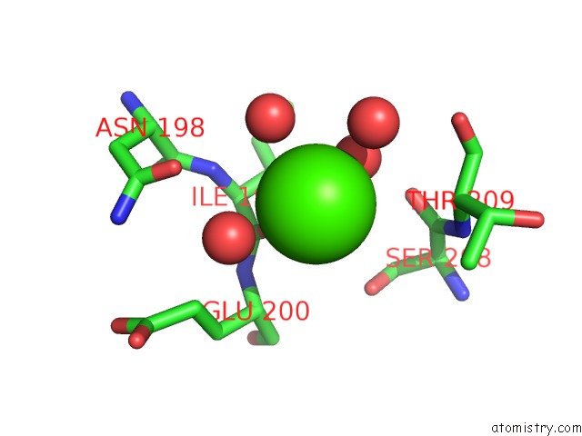
Mono view
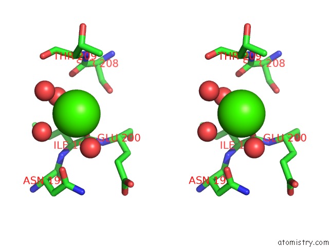
Stereo pair view
Mono view
Stereo pair view
A full contact list of Calcium with other atoms in the Ca binding
site number 1 of Structural Insights Into the Crispr-Cas-Associated Ribonuclease Activity of Staphylococcus Epidermidis CSM3 within 5.0Å range:
|
Reference:
Y.Q.Zhao,
Y.J.Gu,
X.F.Zhu,
W.Cheng.
Structural Insights Into the Crispr-Cas-Associated Ribonuclease Activity of Staphylococcus Epidermidis CSM3 and CSM6 To Be Published.
Page generated: Wed Jul 9 11:58:40 2025
Last articles
Ca in 7G68Ca in 7G65
Ca in 7G64
Ca in 7G67
Ca in 7G66
Ca in 7G63
Ca in 7G61
Ca in 7G62
Ca in 7G60
Ca in 7G5Z