Calcium »
PDB 5yjd-5z4p »
5yjh »
Calcium in PDB 5yjh: Structural Insights Into Periostin Functions
Protein crystallography data
The structure of Structural Insights Into Periostin Functions, PDB code: 5yjh
was solved by
H.Liu,
J.Liu,
F.Xu,
with X-Ray Crystallography technique. A brief refinement statistics is given in the table below:
| Resolution Low / High (Å) | 44.04 / 2.96 |
| Space group | P 61 2 2 |
| Cell size a, b, c (Å), α, β, γ (°) | 105.529, 105.529, 330.342, 90.00, 90.00, 120.00 |
| R / Rfree (%) | 21.3 / 24.2 |
Other elements in 5yjh:
The structure of Structural Insights Into Periostin Functions also contains other interesting chemical elements:
| Magnesium | (Mg) | 9 atoms |
| Zinc | (Zn) | 1 atom |
| Chlorine | (Cl) | 18 atoms |
| Sodium | (Na) | 1 atom |
Calcium Binding Sites:
The binding sites of Calcium atom in the Structural Insights Into Periostin Functions
(pdb code 5yjh). This binding sites where shown within
5.0 Angstroms radius around Calcium atom.
In total 3 binding sites of Calcium where determined in the Structural Insights Into Periostin Functions, PDB code: 5yjh:
Jump to Calcium binding site number: 1; 2; 3;
In total 3 binding sites of Calcium where determined in the Structural Insights Into Periostin Functions, PDB code: 5yjh:
Jump to Calcium binding site number: 1; 2; 3;
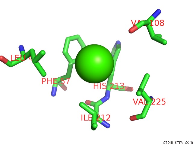
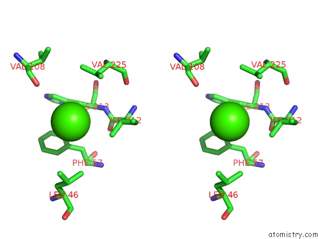
Calcium binding site 1 out of 3 in 5yjh
Go back to
Calcium binding site 1 out
of 3 in the Structural Insights Into Periostin Functions
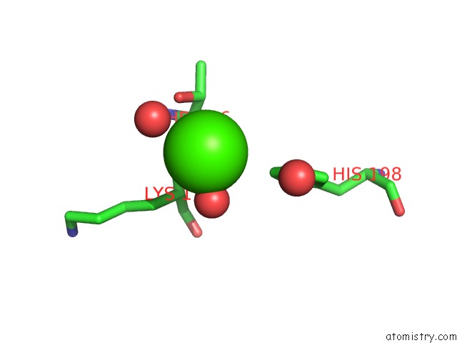
Mono view
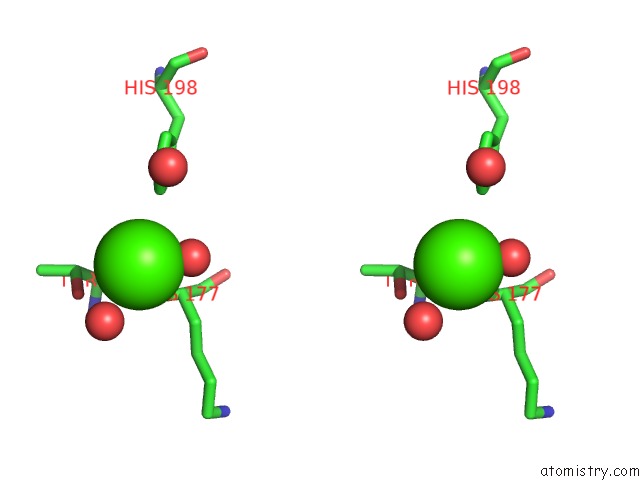
Stereo pair view
Mono view
Stereo pair view
A full contact list of Calcium with other atoms in the Ca binding
site number 1 of Structural Insights Into Periostin Functions within 5.0Å range:
|
Calcium binding site 2 out of 3 in 5yjh
Go back to
Calcium binding site 2 out
of 3 in the Structural Insights Into Periostin Functions
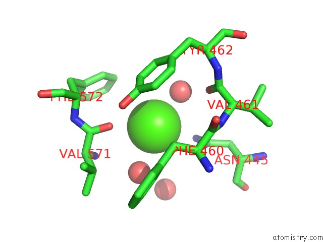
Mono view
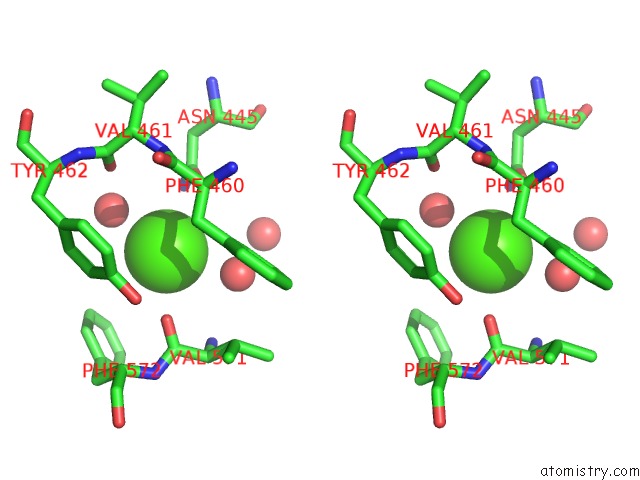
Stereo pair view
Mono view
Stereo pair view
A full contact list of Calcium with other atoms in the Ca binding
site number 2 of Structural Insights Into Periostin Functions within 5.0Å range:
|
Calcium binding site 3 out of 3 in 5yjh
Go back to
Calcium binding site 3 out
of 3 in the Structural Insights Into Periostin Functions
Mono view
Stereo pair view
Mono view
Stereo pair view
A full contact list of Calcium with other atoms in the Ca binding
site number 3 of Structural Insights Into Periostin Functions within 5.0Å range:
|
Reference:
J.Liu,
J.Zhang,
F.Xu,
Z.Lin,
Z.Li,
H.Liu.
Structural Characterizations of Human Periostin Dimerization and Cysteinylation. Febs Lett. V. 592 1789 2018.
ISSN: ISSN 1873-3468
PubMed: 29754429
DOI: 10.1002/1873-3468.13091
Page generated: Wed Jul 9 11:58:40 2025
ISSN: ISSN 1873-3468
PubMed: 29754429
DOI: 10.1002/1873-3468.13091
Last articles
Cl in 5QJMCl in 5QJL
Cl in 5QJB
Cl in 5QJK
Cl in 5QJJ
Cl in 5QJI
Cl in 5QJH
Cl in 5QJG
Cl in 5QJF
Cl in 5QJE