Calcium »
PDB 5yjd-5z4p »
5ynd »
Calcium in PDB 5ynd: Crystal Structure of Pullulanase From Klebsiella Pneumoniae Complex at 1 Mm Gamma-Cyclodextrin
Protein crystallography data
The structure of Crystal Structure of Pullulanase From Klebsiella Pneumoniae Complex at 1 Mm Gamma-Cyclodextrin, PDB code: 5ynd
was solved by
N.Saka,
H.Iwamoto,
N.Takahashi,
K.Mizutani,
B.Mikami,
with X-Ray Crystallography technique. A brief refinement statistics is given in the table below:
| Resolution Low / High (Å) | 44.39 / 2.23 |
| Space group | P 43 21 2 |
| Cell size a, b, c (Å), α, β, γ (°) | 88.773, 88.773, 298.379, 90.00, 90.00, 90.00 |
| R / Rfree (%) | 19.1 / 24.3 |
Calcium Binding Sites:
The binding sites of Calcium atom in the Crystal Structure of Pullulanase From Klebsiella Pneumoniae Complex at 1 Mm Gamma-Cyclodextrin
(pdb code 5ynd). This binding sites where shown within
5.0 Angstroms radius around Calcium atom.
In total 5 binding sites of Calcium where determined in the Crystal Structure of Pullulanase From Klebsiella Pneumoniae Complex at 1 Mm Gamma-Cyclodextrin, PDB code: 5ynd:
Jump to Calcium binding site number: 1; 2; 3; 4; 5;
In total 5 binding sites of Calcium where determined in the Crystal Structure of Pullulanase From Klebsiella Pneumoniae Complex at 1 Mm Gamma-Cyclodextrin, PDB code: 5ynd:
Jump to Calcium binding site number: 1; 2; 3; 4; 5;
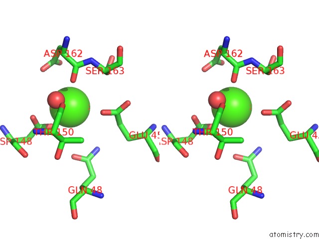
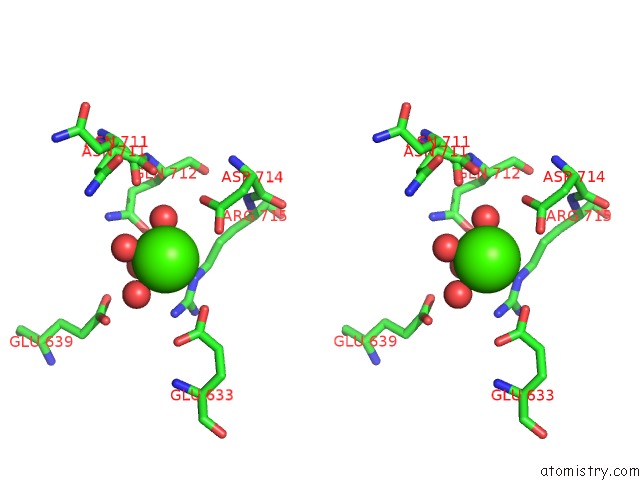
Calcium binding site 1 out of 5 in 5ynd
Go back to
Calcium binding site 1 out
of 5 in the Crystal Structure of Pullulanase From Klebsiella Pneumoniae Complex at 1 Mm Gamma-Cyclodextrin
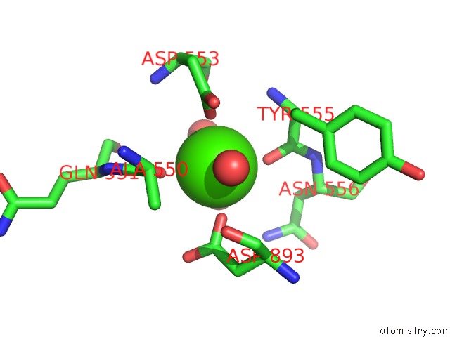
Mono view

Stereo pair view
Mono view
Stereo pair view
A full contact list of Calcium with other atoms in the Ca binding
site number 1 of Crystal Structure of Pullulanase From Klebsiella Pneumoniae Complex at 1 Mm Gamma-Cyclodextrin within 5.0Å range:
|
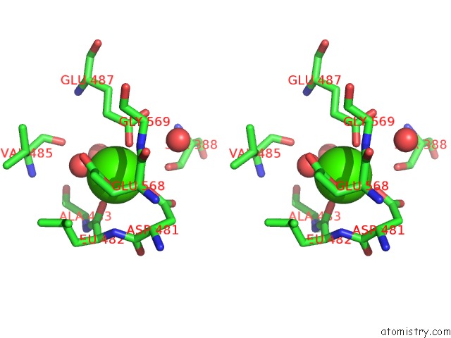
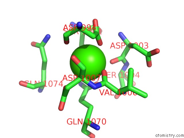
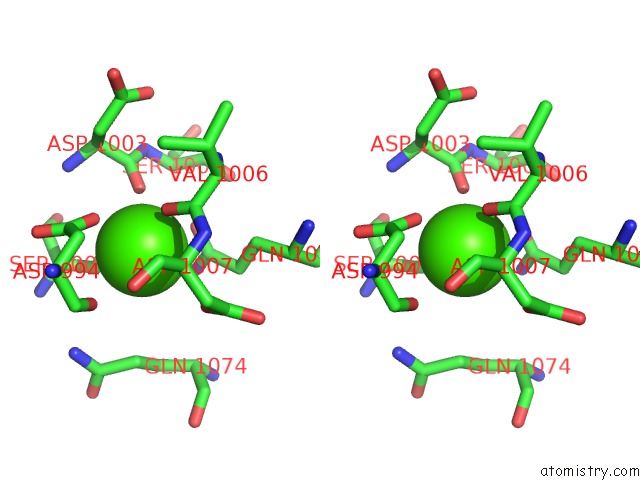
Calcium binding site 2 out of 5 in 5ynd
Go back to
Calcium binding site 2 out
of 5 in the Crystal Structure of Pullulanase From Klebsiella Pneumoniae Complex at 1 Mm Gamma-Cyclodextrin
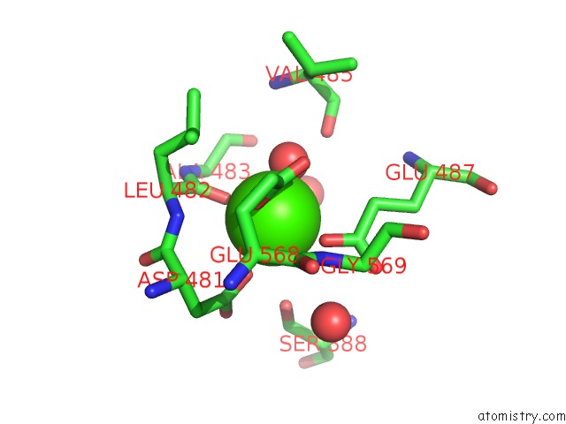
Mono view
Stereo pair view
Mono view
Stereo pair view
A full contact list of Calcium with other atoms in the Ca binding
site number 2 of Crystal Structure of Pullulanase From Klebsiella Pneumoniae Complex at 1 Mm Gamma-Cyclodextrin within 5.0Å range:
|
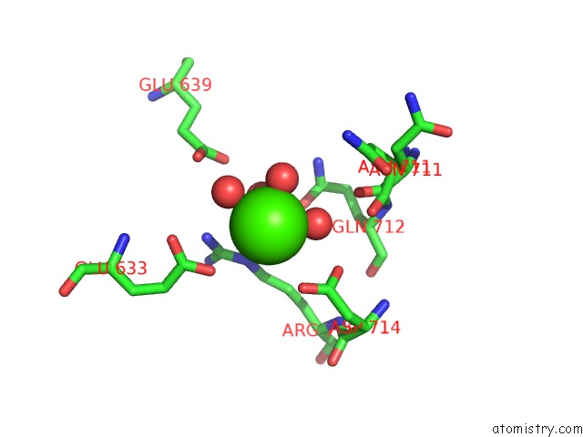
Calcium binding site 3 out of 5 in 5ynd
Go back to
Calcium binding site 3 out
of 5 in the Crystal Structure of Pullulanase From Klebsiella Pneumoniae Complex at 1 Mm Gamma-Cyclodextrin
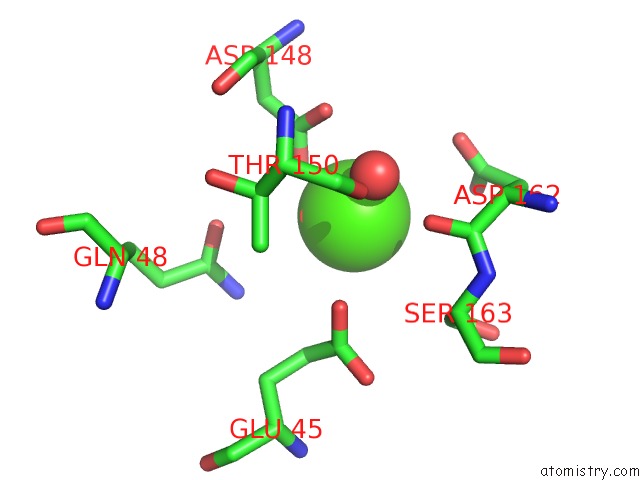
Mono view
Stereo pair view
Mono view
Stereo pair view
A full contact list of Calcium with other atoms in the Ca binding
site number 3 of Crystal Structure of Pullulanase From Klebsiella Pneumoniae Complex at 1 Mm Gamma-Cyclodextrin within 5.0Å range:
|
Calcium binding site 4 out of 5 in 5ynd
Go back to
Calcium binding site 4 out
of 5 in the Crystal Structure of Pullulanase From Klebsiella Pneumoniae Complex at 1 Mm Gamma-Cyclodextrin
Mono view
Stereo pair view
Mono view
Stereo pair view
A full contact list of Calcium with other atoms in the Ca binding
site number 4 of Crystal Structure of Pullulanase From Klebsiella Pneumoniae Complex at 1 Mm Gamma-Cyclodextrin within 5.0Å range:
|
Calcium binding site 5 out of 5 in 5ynd
Go back to
Calcium binding site 5 out
of 5 in the Crystal Structure of Pullulanase From Klebsiella Pneumoniae Complex at 1 Mm Gamma-Cyclodextrin
Mono view
Stereo pair view
Mono view
Stereo pair view
A full contact list of Calcium with other atoms in the Ca binding
site number 5 of Crystal Structure of Pullulanase From Klebsiella Pneumoniae Complex at 1 Mm Gamma-Cyclodextrin within 5.0Å range:
|
Reference:
N.Saka,
H.Iwamoto,
D.Malle,
N.Takahashi,
K.Mizutani,
B.Mikami.
Elucidation of the Mechanism of Interaction Between Klebsiella Pneumoniae Pullulanase and Cyclodextrin Acta Crystallogr D Struct V. 74 1115 2018BIOL.
ISSN: ISSN 2059-7983
PubMed: 30387770
DOI: 10.1107/S2059798318014523
Page generated: Mon Jul 15 15:30:30 2024
ISSN: ISSN 2059-7983
PubMed: 30387770
DOI: 10.1107/S2059798318014523
Last articles
Zn in 9J0NZn in 9J0O
Zn in 9J0P
Zn in 9FJX
Zn in 9EKB
Zn in 9C0F
Zn in 9CAH
Zn in 9CH0
Zn in 9CH3
Zn in 9CH1