Calcium »
PDB 5yjd-5z4p »
5ypu »
Calcium in PDB 5ypu: Crystal Structure of An Actin Monomer in Complex with the Nucleator Cordon-Bleu MET72NLE WH2-Motif Peptide
Protein crystallography data
The structure of Crystal Structure of An Actin Monomer in Complex with the Nucleator Cordon-Bleu MET72NLE WH2-Motif Peptide, PDB code: 5ypu
was solved by
C.P.M.Scipion,
J.Wongsantichon,
F.J.Ferrer,
T.Y.Yuen,
R.C.Robinson,
with X-Ray Crystallography technique. A brief refinement statistics is given in the table below:
| Resolution Low / High (Å) | 29.82 / 2.00 |
| Space group | C 1 2 1 |
| Cell size a, b, c (Å), α, β, γ (°) | 174.861, 40.832, 109.000, 90.00, 101.76, 90.00 |
| R / Rfree (%) | 19.3 / 24 |
Calcium Binding Sites:
The binding sites of Calcium atom in the Crystal Structure of An Actin Monomer in Complex with the Nucleator Cordon-Bleu MET72NLE WH2-Motif Peptide
(pdb code 5ypu). This binding sites where shown within
5.0 Angstroms radius around Calcium atom.
In total 7 binding sites of Calcium where determined in the Crystal Structure of An Actin Monomer in Complex with the Nucleator Cordon-Bleu MET72NLE WH2-Motif Peptide, PDB code: 5ypu:
Jump to Calcium binding site number: 1; 2; 3; 4; 5; 6; 7;
In total 7 binding sites of Calcium where determined in the Crystal Structure of An Actin Monomer in Complex with the Nucleator Cordon-Bleu MET72NLE WH2-Motif Peptide, PDB code: 5ypu:
Jump to Calcium binding site number: 1; 2; 3; 4; 5; 6; 7;
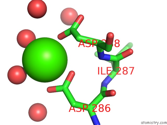
Calcium binding site 1 out of 7 in 5ypu
Go back to
Calcium binding site 1 out
of 7 in the Crystal Structure of An Actin Monomer in Complex with the Nucleator Cordon-Bleu MET72NLE WH2-Motif Peptide

Mono view

Stereo pair view
Mono view
Stereo pair view
A full contact list of Calcium with other atoms in the Ca binding
site number 1 of Crystal Structure of An Actin Monomer in Complex with the Nucleator Cordon-Bleu MET72NLE WH2-Motif Peptide within 5.0Å range:
|
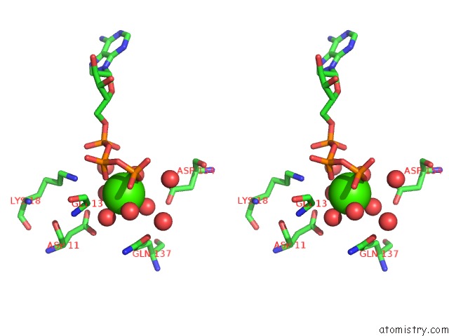
Calcium binding site 2 out of 7 in 5ypu
Go back to
Calcium binding site 2 out
of 7 in the Crystal Structure of An Actin Monomer in Complex with the Nucleator Cordon-Bleu MET72NLE WH2-Motif Peptide
Mono view

Stereo pair view
Mono view
Stereo pair view
A full contact list of Calcium with other atoms in the Ca binding
site number 2 of Crystal Structure of An Actin Monomer in Complex with the Nucleator Cordon-Bleu MET72NLE WH2-Motif Peptide within 5.0Å range:
|
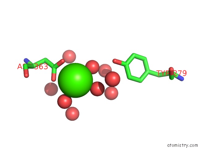
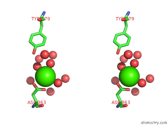
Calcium binding site 3 out of 7 in 5ypu
Go back to
Calcium binding site 3 out
of 7 in the Crystal Structure of An Actin Monomer in Complex with the Nucleator Cordon-Bleu MET72NLE WH2-Motif Peptide

Mono view

Stereo pair view
Mono view
Stereo pair view
A full contact list of Calcium with other atoms in the Ca binding
site number 3 of Crystal Structure of An Actin Monomer in Complex with the Nucleator Cordon-Bleu MET72NLE WH2-Motif Peptide within 5.0Å range:
|
Calcium binding site 4 out of 7 in 5ypu
Go back to
Calcium binding site 4 out
of 7 in the Crystal Structure of An Actin Monomer in Complex with the Nucleator Cordon-Bleu MET72NLE WH2-Motif Peptide

Mono view
Stereo pair view
Mono view
Stereo pair view
A full contact list of Calcium with other atoms in the Ca binding
site number 4 of Crystal Structure of An Actin Monomer in Complex with the Nucleator Cordon-Bleu MET72NLE WH2-Motif Peptide within 5.0Å range:
|
Calcium binding site 5 out of 7 in 5ypu
Go back to
Calcium binding site 5 out
of 7 in the Crystal Structure of An Actin Monomer in Complex with the Nucleator Cordon-Bleu MET72NLE WH2-Motif Peptide

Mono view

Stereo pair view
Mono view
Stereo pair view
A full contact list of Calcium with other atoms in the Ca binding
site number 5 of Crystal Structure of An Actin Monomer in Complex with the Nucleator Cordon-Bleu MET72NLE WH2-Motif Peptide within 5.0Å range:
|
Calcium binding site 6 out of 7 in 5ypu
Go back to
Calcium binding site 6 out
of 7 in the Crystal Structure of An Actin Monomer in Complex with the Nucleator Cordon-Bleu MET72NLE WH2-Motif Peptide
Mono view
Stereo pair view
Mono view
Stereo pair view
A full contact list of Calcium with other atoms in the Ca binding
site number 6 of Crystal Structure of An Actin Monomer in Complex with the Nucleator Cordon-Bleu MET72NLE WH2-Motif Peptide within 5.0Å range:
|
Calcium binding site 7 out of 7 in 5ypu
Go back to
Calcium binding site 7 out
of 7 in the Crystal Structure of An Actin Monomer in Complex with the Nucleator Cordon-Bleu MET72NLE WH2-Motif Peptide

Mono view

Stereo pair view
Mono view
Stereo pair view
A full contact list of Calcium with other atoms in the Ca binding
site number 7 of Crystal Structure of An Actin Monomer in Complex with the Nucleator Cordon-Bleu MET72NLE WH2-Motif Peptide within 5.0Å range:
|
Reference:
C.P.M.Scipion,
U.Ghoshdastider,
F.J.Ferrer,
T.Y.Yuen,
J.Wongsantichon,
R.C.Robinson.
Structural Evidence For the Roles of Divalent Cations in Actin Polymerization and Activation of Atp Hydrolysis Proc. Natl. Acad. Sci. V. 115 10345 2018U.S.A..
ISSN: ESSN 1091-6490
PubMed: 30254171
DOI: 10.1073/PNAS.1806394115
Page generated: Wed Jul 9 12:02:43 2025
ISSN: ESSN 1091-6490
PubMed: 30254171
DOI: 10.1073/PNAS.1806394115
Last articles
Cl in 5QFJCl in 5QF2
Cl in 5QF7
Cl in 5QED
Cl in 5QFA
Cl in 5QEW
Cl in 5QEJ
Cl in 5QEV
Cl in 5QES
Cl in 5QEU