Calcium »
PDB 5z4u-5zxh »
5zce »
Calcium in PDB 5zce: Crystal Structure of Alpha-Glucosidase in Complex with Maltotetraose
Protein crystallography data
The structure of Crystal Structure of Alpha-Glucosidase in Complex with Maltotetraose, PDB code: 5zce
was solved by
K.Kato,
W.Saburi,
M.Yao,
with X-Ray Crystallography technique. A brief refinement statistics is given in the table below:
| Resolution Low / High (Å) | 46.10 / 1.56 |
| Space group | P 21 21 21 |
| Cell size a, b, c (Å), α, β, γ (°) | 54.989, 84.549, 128.715, 90.00, 90.00, 90.00 |
| R / Rfree (%) | 17.2 / 19.1 |
Calcium Binding Sites:
The binding sites of Calcium atom in the Crystal Structure of Alpha-Glucosidase in Complex with Maltotetraose
(pdb code 5zce). This binding sites where shown within
5.0 Angstroms radius around Calcium atom.
In total 3 binding sites of Calcium where determined in the Crystal Structure of Alpha-Glucosidase in Complex with Maltotetraose, PDB code: 5zce:
Jump to Calcium binding site number: 1; 2; 3;
In total 3 binding sites of Calcium where determined in the Crystal Structure of Alpha-Glucosidase in Complex with Maltotetraose, PDB code: 5zce:
Jump to Calcium binding site number: 1; 2; 3;
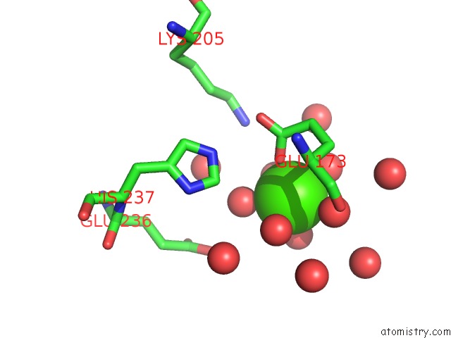
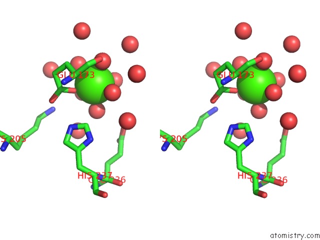
Calcium binding site 1 out of 3 in 5zce
Go back to
Calcium binding site 1 out
of 3 in the Crystal Structure of Alpha-Glucosidase in Complex with Maltotetraose
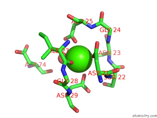
Mono view
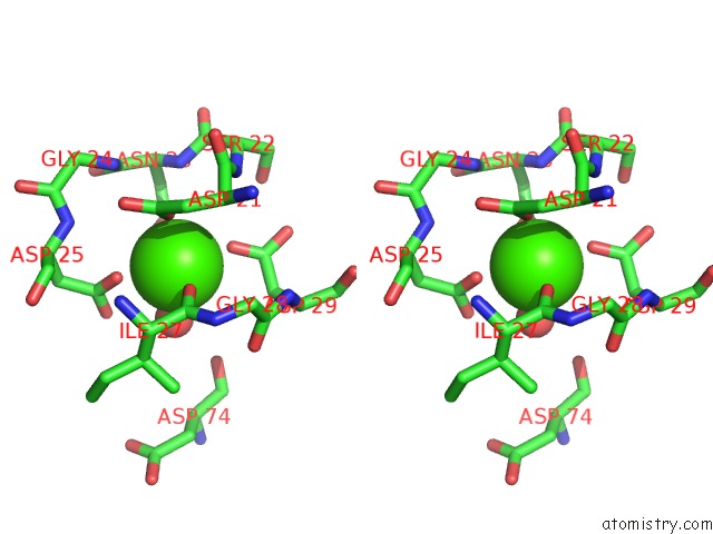
Stereo pair view
Mono view
Stereo pair view
A full contact list of Calcium with other atoms in the Ca binding
site number 1 of Crystal Structure of Alpha-Glucosidase in Complex with Maltotetraose within 5.0Å range:
|
Calcium binding site 2 out of 3 in 5zce
Go back to
Calcium binding site 2 out
of 3 in the Crystal Structure of Alpha-Glucosidase in Complex with Maltotetraose
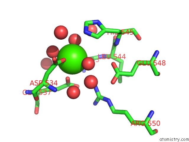
Mono view
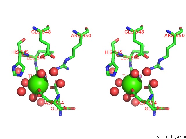
Stereo pair view
Mono view
Stereo pair view
A full contact list of Calcium with other atoms in the Ca binding
site number 2 of Crystal Structure of Alpha-Glucosidase in Complex with Maltotetraose within 5.0Å range:
|
Calcium binding site 3 out of 3 in 5zce
Go back to
Calcium binding site 3 out
of 3 in the Crystal Structure of Alpha-Glucosidase in Complex with Maltotetraose
Mono view
Stereo pair view
Mono view
Stereo pair view
A full contact list of Calcium with other atoms in the Ca binding
site number 3 of Crystal Structure of Alpha-Glucosidase in Complex with Maltotetraose within 5.0Å range:
|
Reference:
W.Auiewiriyanukul,
W.Saburi,
K.Kato,
M.Yao,
H.Mori.
Function and Structure of GH13_31 Alpha-Glucosidase with High Alpha-(1→4)-Glucosidic Linkage Specificity and Transglucosylation Activity. Febs Lett. V. 592 2268 2018.
ISSN: ISSN 1873-3468
PubMed: 29870070
DOI: 10.1002/1873-3468.13126
Page generated: Mon Jul 15 15:49:08 2024
ISSN: ISSN 1873-3468
PubMed: 29870070
DOI: 10.1002/1873-3468.13126
Last articles
Zn in 9J0NZn in 9J0O
Zn in 9J0P
Zn in 9FJX
Zn in 9EKB
Zn in 9C0F
Zn in 9CAH
Zn in 9CH0
Zn in 9CH3
Zn in 9CH1