Calcium »
PDB 5z4u-5zxh »
5zdm »
Calcium in PDB 5zdm: The Ligand-Free Structure of Fomd
Protein crystallography data
The structure of The Ligand-Free Structure of Fomd, PDB code: 5zdm
was solved by
S.Sato,
A.Miyanaga,
F.Kudo,
T.Eguchi,
with X-Ray Crystallography technique. A brief refinement statistics is given in the table below:
| Resolution Low / High (Å) | 49.25 / 1.38 |
| Space group | P 1 21 1 |
| Cell size a, b, c (Å), α, β, γ (°) | 40.474, 46.215, 52.504, 90.00, 110.27, 90.00 |
| R / Rfree (%) | 16.4 / 19.2 |
Calcium Binding Sites:
The binding sites of Calcium atom in the The Ligand-Free Structure of Fomd
(pdb code 5zdm). This binding sites where shown within
5.0 Angstroms radius around Calcium atom.
In total 2 binding sites of Calcium where determined in the The Ligand-Free Structure of Fomd, PDB code: 5zdm:
Jump to Calcium binding site number: 1; 2;
In total 2 binding sites of Calcium where determined in the The Ligand-Free Structure of Fomd, PDB code: 5zdm:
Jump to Calcium binding site number: 1; 2;
Calcium binding site 1 out of 2 in 5zdm
Go back to
Calcium binding site 1 out
of 2 in the The Ligand-Free Structure of Fomd
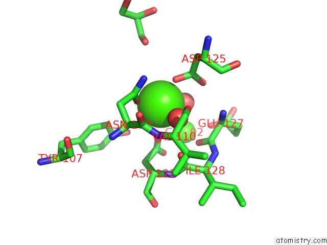
Mono view
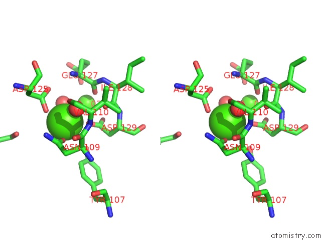
Stereo pair view
Mono view
Stereo pair view
A full contact list of Calcium with other atoms in the Ca binding
site number 1 of The Ligand-Free Structure of Fomd within 5.0Å range:
|
Calcium binding site 2 out of 2 in 5zdm
Go back to
Calcium binding site 2 out
of 2 in the The Ligand-Free Structure of Fomd
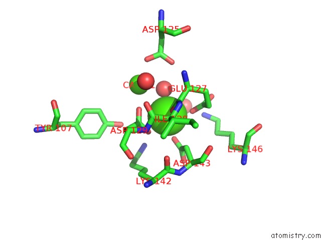
Mono view
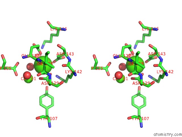
Stereo pair view
Mono view
Stereo pair view
A full contact list of Calcium with other atoms in the Ca binding
site number 2 of The Ligand-Free Structure of Fomd within 5.0Å range:
|
Reference:
S.Sato,
A.Miyanaga,
S.Y.Kim,
T.Kuzuyama,
F.Kudo,
T.Eguchi.
Biochemical and Structural Analysis of Fomd That Catalyzes the Hydrolysis of Cytidylyl ( S)-2-Hydroxypropylphosphonate in Fosfomycin Biosynthesis. Biochemistry V. 57 4858 2018.
ISSN: ISSN 1520-4995
PubMed: 30010320
DOI: 10.1021/ACS.BIOCHEM.8B00690
Page generated: Mon Jul 15 15:49:21 2024
ISSN: ISSN 1520-4995
PubMed: 30010320
DOI: 10.1021/ACS.BIOCHEM.8B00690
Last articles
Zn in 9J0NZn in 9J0O
Zn in 9J0P
Zn in 9FJX
Zn in 9EKB
Zn in 9C0F
Zn in 9CAH
Zn in 9CH0
Zn in 9CH3
Zn in 9CH1