Calcium »
PDB 5z4u-5zxh »
5zno »
Calcium in PDB 5zno: Crystal Structure of Pet-Degrading Cutinase CUT190 S176A/S226P/R228S/ Mutant in Ca(2+)-Bound State
Enzymatic activity of Crystal Structure of Pet-Degrading Cutinase CUT190 S176A/S226P/R228S/ Mutant in Ca(2+)-Bound State
All present enzymatic activity of Crystal Structure of Pet-Degrading Cutinase CUT190 S176A/S226P/R228S/ Mutant in Ca(2+)-Bound State:
3.1.1.74;
3.1.1.74;
Protein crystallography data
The structure of Crystal Structure of Pet-Degrading Cutinase CUT190 S176A/S226P/R228S/ Mutant in Ca(2+)-Bound State, PDB code: 5zno
was solved by
N.Numoto,
S.Inaba,
Y.Yamagami,
N.Kamiya,
G.J.Bekker,
K.Ishii,
S.Uchiyama,
F.Kawai,
N.Ito,
M.Oda,
with X-Ray Crystallography technique. A brief refinement statistics is given in the table below:
| Resolution Low / High (Å) | 47.72 / 1.60 |
| Space group | C 1 2 1 |
| Cell size a, b, c (Å), α, β, γ (°) | 123.705, 49.732, 95.655, 90.00, 99.59, 90.00 |
| R / Rfree (%) | 17.9 / 22.2 |
Calcium Binding Sites:
The binding sites of Calcium atom in the Crystal Structure of Pet-Degrading Cutinase CUT190 S176A/S226P/R228S/ Mutant in Ca(2+)-Bound State
(pdb code 5zno). This binding sites where shown within
5.0 Angstroms radius around Calcium atom.
In total 7 binding sites of Calcium where determined in the Crystal Structure of Pet-Degrading Cutinase CUT190 S176A/S226P/R228S/ Mutant in Ca(2+)-Bound State, PDB code: 5zno:
Jump to Calcium binding site number: 1; 2; 3; 4; 5; 6; 7;
In total 7 binding sites of Calcium where determined in the Crystal Structure of Pet-Degrading Cutinase CUT190 S176A/S226P/R228S/ Mutant in Ca(2+)-Bound State, PDB code: 5zno:
Jump to Calcium binding site number: 1; 2; 3; 4; 5; 6; 7;
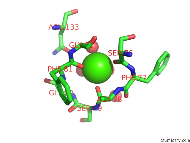
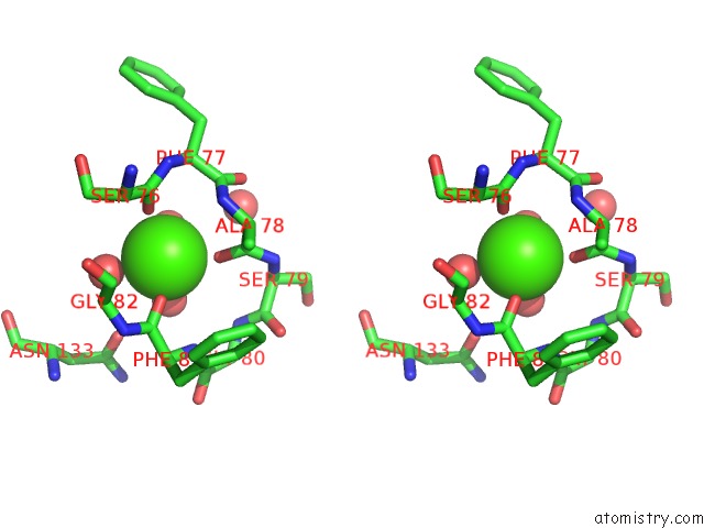
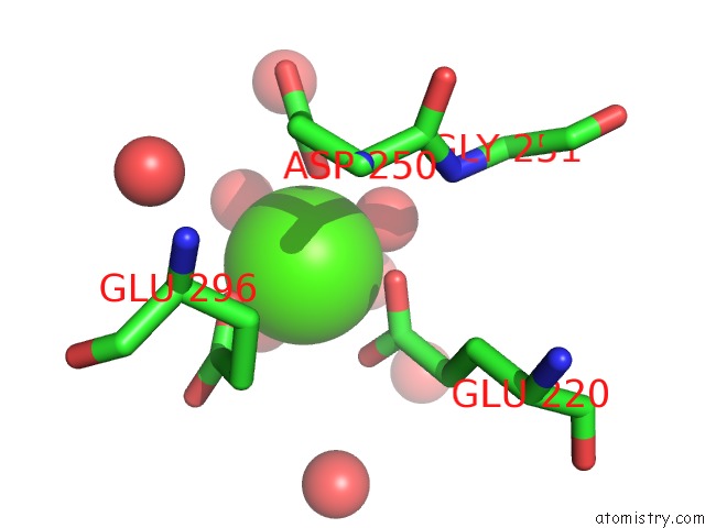
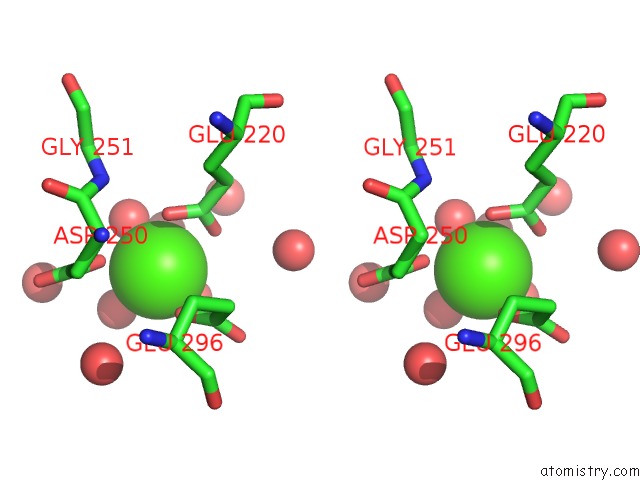
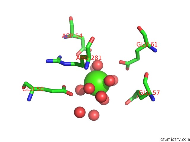
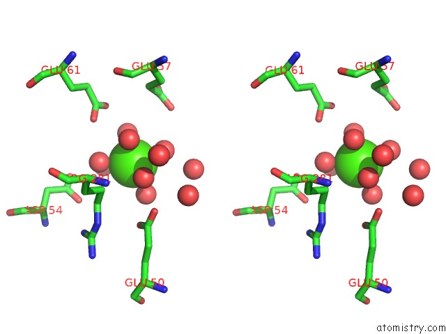
Calcium binding site 1 out of 7 in 5zno
Go back to
Calcium binding site 1 out
of 7 in the Crystal Structure of Pet-Degrading Cutinase CUT190 S176A/S226P/R228S/ Mutant in Ca(2+)-Bound State
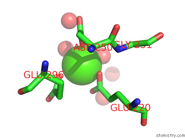
Mono view
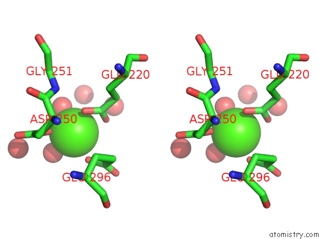
Stereo pair view
Mono view
Stereo pair view
A full contact list of Calcium with other atoms in the Ca binding
site number 1 of Crystal Structure of Pet-Degrading Cutinase CUT190 S176A/S226P/R228S/ Mutant in Ca(2+)-Bound State within 5.0Å range:
|
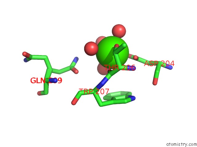
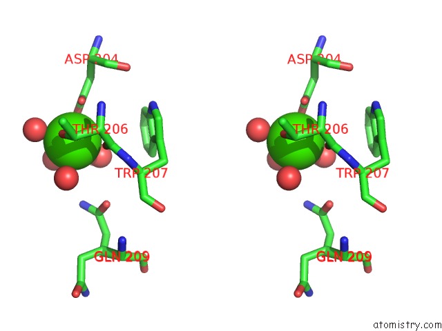
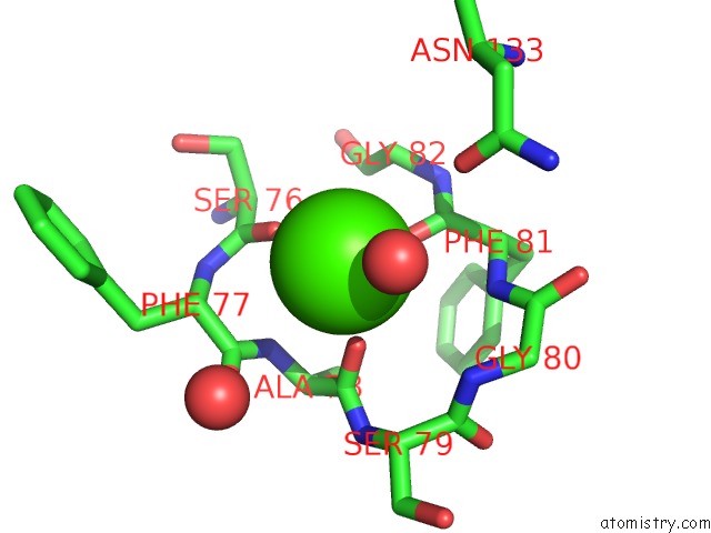
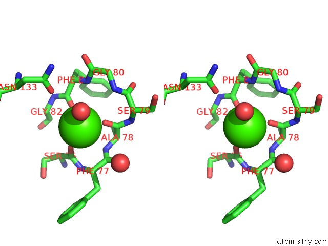
Calcium binding site 2 out of 7 in 5zno
Go back to
Calcium binding site 2 out
of 7 in the Crystal Structure of Pet-Degrading Cutinase CUT190 S176A/S226P/R228S/ Mutant in Ca(2+)-Bound State
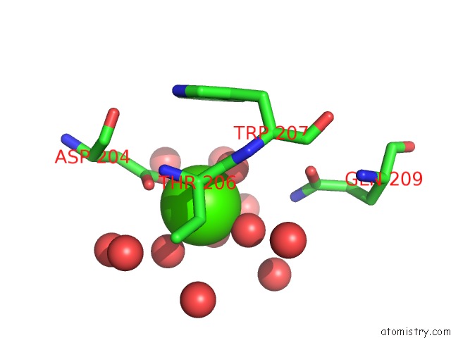
Mono view
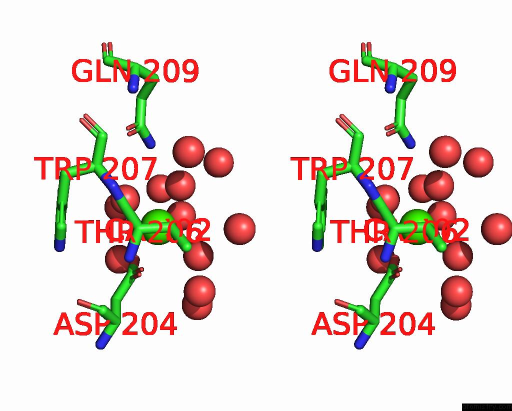
Stereo pair view
Mono view
Stereo pair view
A full contact list of Calcium with other atoms in the Ca binding
site number 2 of Crystal Structure of Pet-Degrading Cutinase CUT190 S176A/S226P/R228S/ Mutant in Ca(2+)-Bound State within 5.0Å range:
|
Calcium binding site 3 out of 7 in 5zno
Go back to
Calcium binding site 3 out
of 7 in the Crystal Structure of Pet-Degrading Cutinase CUT190 S176A/S226P/R228S/ Mutant in Ca(2+)-Bound State
Mono view
Stereo pair view
Mono view
Stereo pair view
A full contact list of Calcium with other atoms in the Ca binding
site number 3 of Crystal Structure of Pet-Degrading Cutinase CUT190 S176A/S226P/R228S/ Mutant in Ca(2+)-Bound State within 5.0Å range:
|
Calcium binding site 4 out of 7 in 5zno
Go back to
Calcium binding site 4 out
of 7 in the Crystal Structure of Pet-Degrading Cutinase CUT190 S176A/S226P/R228S/ Mutant in Ca(2+)-Bound State
Mono view
Stereo pair view
Mono view
Stereo pair view
A full contact list of Calcium with other atoms in the Ca binding
site number 4 of Crystal Structure of Pet-Degrading Cutinase CUT190 S176A/S226P/R228S/ Mutant in Ca(2+)-Bound State within 5.0Å range:
|
Calcium binding site 5 out of 7 in 5zno
Go back to
Calcium binding site 5 out
of 7 in the Crystal Structure of Pet-Degrading Cutinase CUT190 S176A/S226P/R228S/ Mutant in Ca(2+)-Bound State
Mono view
Stereo pair view
Mono view
Stereo pair view
A full contact list of Calcium with other atoms in the Ca binding
site number 5 of Crystal Structure of Pet-Degrading Cutinase CUT190 S176A/S226P/R228S/ Mutant in Ca(2+)-Bound State within 5.0Å range:
|
Calcium binding site 6 out of 7 in 5zno
Go back to
Calcium binding site 6 out
of 7 in the Crystal Structure of Pet-Degrading Cutinase CUT190 S176A/S226P/R228S/ Mutant in Ca(2+)-Bound State
Mono view
Stereo pair view
Mono view
Stereo pair view
A full contact list of Calcium with other atoms in the Ca binding
site number 6 of Crystal Structure of Pet-Degrading Cutinase CUT190 S176A/S226P/R228S/ Mutant in Ca(2+)-Bound State within 5.0Å range:
|
Calcium binding site 7 out of 7 in 5zno
Go back to
Calcium binding site 7 out
of 7 in the Crystal Structure of Pet-Degrading Cutinase CUT190 S176A/S226P/R228S/ Mutant in Ca(2+)-Bound State
Mono view
Stereo pair view
Mono view
Stereo pair view
A full contact list of Calcium with other atoms in the Ca binding
site number 7 of Crystal Structure of Pet-Degrading Cutinase CUT190 S176A/S226P/R228S/ Mutant in Ca(2+)-Bound State within 5.0Å range:
|
Reference:
N.Numoto,
N.Kamiya,
G.J.Bekker,
Y.Yamagami,
S.Inaba,
K.Ishii,
S.Uchiyama,
F.Kawai,
N.Ito,
M.Oda.
Structural Dynamics of the Pet-Degrading Cutinase-Like Enzyme From Saccharomonospora Viridis AHK190 in Substrate-Bound States Elucidates the CA2+-Driven Catalytic Cycle. Biochemistry V. 57 5289 2018.
ISSN: ISSN 1520-4995
PubMed: 30110540
DOI: 10.1021/ACS.BIOCHEM.8B00624
Page generated: Mon Jul 15 15:52:57 2024
ISSN: ISSN 1520-4995
PubMed: 30110540
DOI: 10.1021/ACS.BIOCHEM.8B00624
Last articles
Zn in 9MJ5Zn in 9HNW
Zn in 9G0L
Zn in 9FNE
Zn in 9DZN
Zn in 9E0I
Zn in 9D32
Zn in 9DAK
Zn in 8ZXC
Zn in 8ZUF