Calcium »
PDB 5zyv-6ahg »
6a8i »
Calcium in PDB 6a8i: Crystal Structure of Endo-Arabinanase Abn-Ts D147N Mutant in Complex with Arabinohexaose
Enzymatic activity of Crystal Structure of Endo-Arabinanase Abn-Ts D147N Mutant in Complex with Arabinohexaose
All present enzymatic activity of Crystal Structure of Endo-Arabinanase Abn-Ts D147N Mutant in Complex with Arabinohexaose:
3.2.1.99;
3.2.1.99;
Protein crystallography data
The structure of Crystal Structure of Endo-Arabinanase Abn-Ts D147N Mutant in Complex with Arabinohexaose, PDB code: 6a8i
was solved by
A.Yamaguchi,
T.Tada,
with X-Ray Crystallography technique. A brief refinement statistics is given in the table below:
| Resolution Low / High (Å) | 50.00 / 1.90 |
| Space group | P 1 21 1 |
| Cell size a, b, c (Å), α, β, γ (°) | 45.809, 92.260, 78.673, 90.00, 91.52, 90.00 |
| R / Rfree (%) | 18.1 / 23.3 |
Calcium Binding Sites:
The binding sites of Calcium atom in the Crystal Structure of Endo-Arabinanase Abn-Ts D147N Mutant in Complex with Arabinohexaose
(pdb code 6a8i). This binding sites where shown within
5.0 Angstroms radius around Calcium atom.
In total 2 binding sites of Calcium where determined in the Crystal Structure of Endo-Arabinanase Abn-Ts D147N Mutant in Complex with Arabinohexaose, PDB code: 6a8i:
Jump to Calcium binding site number: 1; 2;
In total 2 binding sites of Calcium where determined in the Crystal Structure of Endo-Arabinanase Abn-Ts D147N Mutant in Complex with Arabinohexaose, PDB code: 6a8i:
Jump to Calcium binding site number: 1; 2;
Calcium binding site 1 out of 2 in 6a8i
Go back to
Calcium binding site 1 out
of 2 in the Crystal Structure of Endo-Arabinanase Abn-Ts D147N Mutant in Complex with Arabinohexaose

Mono view
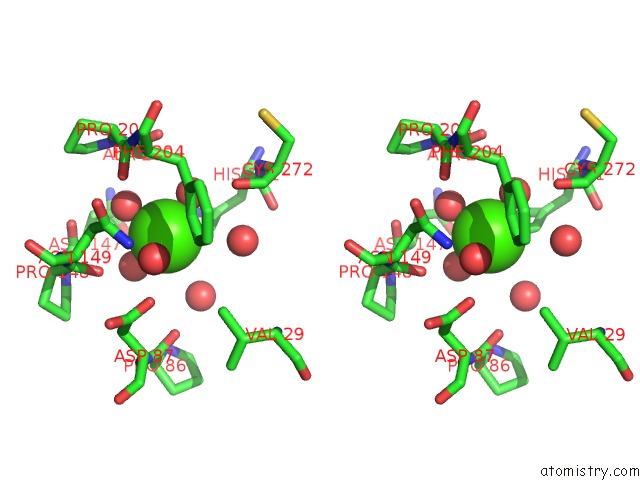
Stereo pair view
Mono view
Stereo pair view
A full contact list of Calcium with other atoms in the Ca binding
site number 1 of Crystal Structure of Endo-Arabinanase Abn-Ts D147N Mutant in Complex with Arabinohexaose within 5.0Å range:
|
Calcium binding site 2 out of 2 in 6a8i
Go back to
Calcium binding site 2 out
of 2 in the Crystal Structure of Endo-Arabinanase Abn-Ts D147N Mutant in Complex with Arabinohexaose

Mono view

Stereo pair view
Mono view
Stereo pair view
A full contact list of Calcium with other atoms in the Ca binding
site number 2 of Crystal Structure of Endo-Arabinanase Abn-Ts D147N Mutant in Complex with Arabinohexaose within 5.0Å range:
|
Reference:
A.Yamaguchi,
Y.Sogabe,
S.Fukuoka,
T.Sakai,
T.Tada.
Structures of Endo-1,5-Alpha-L-Arabinanase Mutants From Bacillus Thermodenitrificans Ts-3 in Complex with Arabino-Oligosaccharides. Acta Crystallogr F Struct V. 74 774 2018BIOL Commun.
ISSN: ESSN 2053-230X
PubMed: 30511671
DOI: 10.1107/S2053230X18015947
Page generated: Wed Jul 9 12:22:02 2025
ISSN: ESSN 2053-230X
PubMed: 30511671
DOI: 10.1107/S2053230X18015947
Last articles
Ca in 7G4FCa in 7G4I
Ca in 7G4G
Ca in 7G4E
Ca in 7G4D
Ca in 7G4B
Ca in 7G4C
Ca in 7G4A
Ca in 7G48
Ca in 7G49