Calcium »
PDB 5zyv-6ahg »
6agk »
Calcium in PDB 6agk: The Structure of Ch-II-77-Tubulin Complex
Protein crystallography data
The structure of The Structure of Ch-II-77-Tubulin Complex, PDB code: 6agk
was solved by
H.Chen,
K.Arnst,
Y.Wang,
D.Miller,
W.Li,
with X-Ray Crystallography technique. A brief refinement statistics is given in the table below:
| Resolution Low / High (Å) | 50.00 / 2.80 |
| Space group | P 21 21 21 |
| Cell size a, b, c (Å), α, β, γ (°) | 105.084, 157.062, 182.897, 90.00, 90.00, 90.00 |
| R / Rfree (%) | 21.1 / 26.4 |
Other elements in 6agk:
The structure of The Structure of Ch-II-77-Tubulin Complex also contains other interesting chemical elements:
| Magnesium | (Mg) | 3 atoms |
Calcium Binding Sites:
The binding sites of Calcium atom in the The Structure of Ch-II-77-Tubulin Complex
(pdb code 6agk). This binding sites where shown within
5.0 Angstroms radius around Calcium atom.
In total 3 binding sites of Calcium where determined in the The Structure of Ch-II-77-Tubulin Complex, PDB code: 6agk:
Jump to Calcium binding site number: 1; 2; 3;
In total 3 binding sites of Calcium where determined in the The Structure of Ch-II-77-Tubulin Complex, PDB code: 6agk:
Jump to Calcium binding site number: 1; 2; 3;
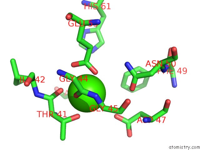
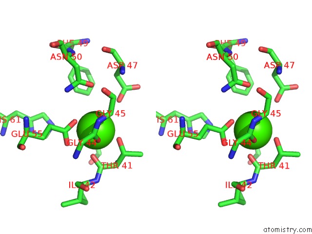
Calcium binding site 1 out of 3 in 6agk
Go back to
Calcium binding site 1 out
of 3 in the The Structure of Ch-II-77-Tubulin Complex
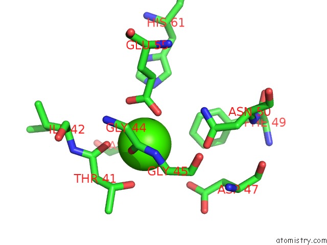
Mono view
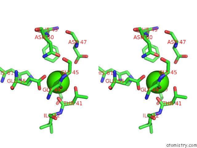
Stereo pair view
Mono view
Stereo pair view
A full contact list of Calcium with other atoms in the Ca binding
site number 1 of The Structure of Ch-II-77-Tubulin Complex within 5.0Å range:
|
Calcium binding site 2 out of 3 in 6agk
Go back to
Calcium binding site 2 out
of 3 in the The Structure of Ch-II-77-Tubulin Complex
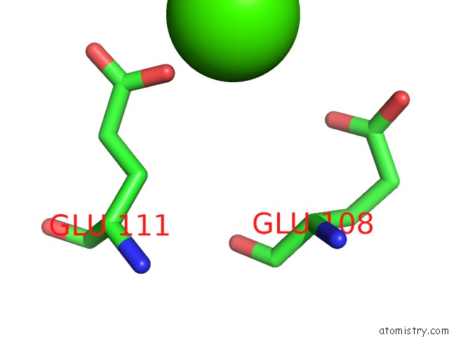
Mono view
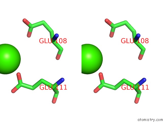
Stereo pair view
Mono view
Stereo pair view
A full contact list of Calcium with other atoms in the Ca binding
site number 2 of The Structure of Ch-II-77-Tubulin Complex within 5.0Å range:
|
Calcium binding site 3 out of 3 in 6agk
Go back to
Calcium binding site 3 out
of 3 in the The Structure of Ch-II-77-Tubulin Complex
Mono view
Stereo pair view
Mono view
Stereo pair view
A full contact list of Calcium with other atoms in the Ca binding
site number 3 of The Structure of Ch-II-77-Tubulin Complex within 5.0Å range:
|
Reference:
H.Chen,
K.Arnst,
Y.Wang,
D.Miller,
W.Li.
The Structure of Ch-II-77-Tubulin Complex To Be Published.
Page generated: Wed Jul 9 12:26:34 2025
Last articles
F in 4HN4F in 4HJX
F in 4HLH
F in 4HL4
F in 4HIQ
F in 4HHZ
F in 4HHY
F in 4HGT
F in 4HEJ
F in 4HGS