Calcium »
PDB 6ai0-6b40 »
6ajb »
Calcium in PDB 6ajb: Crystal Structure of Trypanosoma Brucei Glycosomal Isocitrate Dehydrogenase in Complex with Nadh, Alpha-Ketoglutarate and CA2+
Enzymatic activity of Crystal Structure of Trypanosoma Brucei Glycosomal Isocitrate Dehydrogenase in Complex with Nadh, Alpha-Ketoglutarate and CA2+
All present enzymatic activity of Crystal Structure of Trypanosoma Brucei Glycosomal Isocitrate Dehydrogenase in Complex with Nadh, Alpha-Ketoglutarate and CA2+:
1.1.1.42;
1.1.1.42;
Protein crystallography data
The structure of Crystal Structure of Trypanosoma Brucei Glycosomal Isocitrate Dehydrogenase in Complex with Nadh, Alpha-Ketoglutarate and CA2+, PDB code: 6ajb
was solved by
X.Wang,
D.K.Inaoka,
T.Shiba,
E.O.Balogun,
N.Ziebart,
S.Allman,
Y.Watanabe,
T.Nozaki,
M.Boshart,
F.Bringaud,
S.Harada,
K.Kita,
with X-Ray Crystallography technique. A brief refinement statistics is given in the table below:
| Resolution Low / High (Å) | 19.98 / 2.90 |
| Space group | P 1 21 1 |
| Cell size a, b, c (Å), α, β, γ (°) | 120.520, 70.464, 121.929, 90.00, 113.42, 90.00 |
| R / Rfree (%) | 19.2 / 28.4 |
Calcium Binding Sites:
The binding sites of Calcium atom in the Crystal Structure of Trypanosoma Brucei Glycosomal Isocitrate Dehydrogenase in Complex with Nadh, Alpha-Ketoglutarate and CA2+
(pdb code 6ajb). This binding sites where shown within
5.0 Angstroms radius around Calcium atom.
In total 4 binding sites of Calcium where determined in the Crystal Structure of Trypanosoma Brucei Glycosomal Isocitrate Dehydrogenase in Complex with Nadh, Alpha-Ketoglutarate and CA2+, PDB code: 6ajb:
Jump to Calcium binding site number: 1; 2; 3; 4;
In total 4 binding sites of Calcium where determined in the Crystal Structure of Trypanosoma Brucei Glycosomal Isocitrate Dehydrogenase in Complex with Nadh, Alpha-Ketoglutarate and CA2+, PDB code: 6ajb:
Jump to Calcium binding site number: 1; 2; 3; 4;
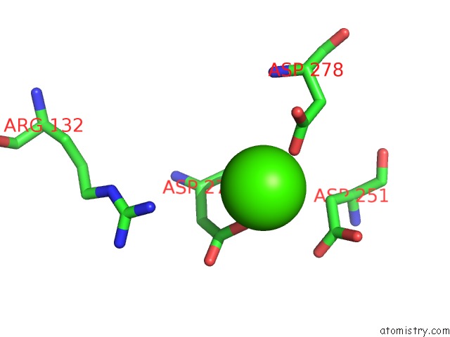
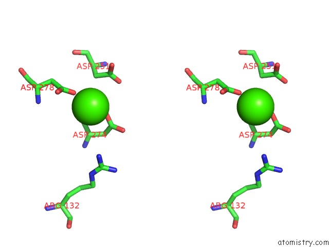
Calcium binding site 1 out of 4 in 6ajb
Go back to
Calcium binding site 1 out
of 4 in the Crystal Structure of Trypanosoma Brucei Glycosomal Isocitrate Dehydrogenase in Complex with Nadh, Alpha-Ketoglutarate and CA2+
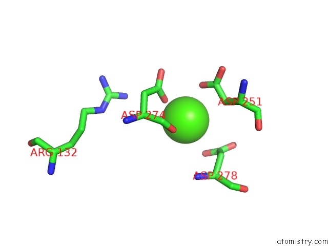
Mono view
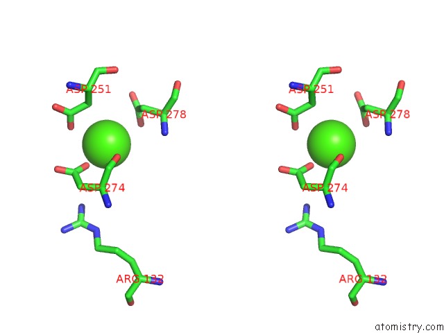
Stereo pair view
Mono view
Stereo pair view
A full contact list of Calcium with other atoms in the Ca binding
site number 1 of Crystal Structure of Trypanosoma Brucei Glycosomal Isocitrate Dehydrogenase in Complex with Nadh, Alpha-Ketoglutarate and CA2+ within 5.0Å range:
|
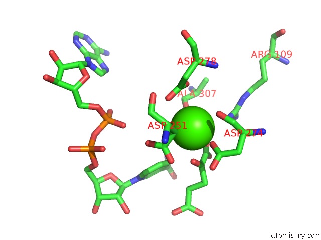
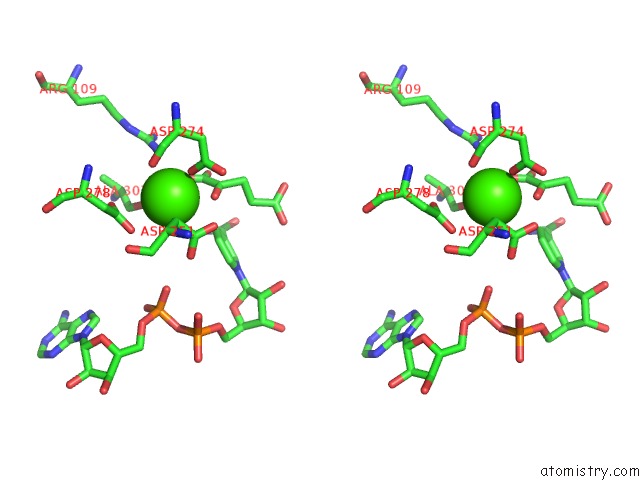
Calcium binding site 2 out of 4 in 6ajb
Go back to
Calcium binding site 2 out
of 4 in the Crystal Structure of Trypanosoma Brucei Glycosomal Isocitrate Dehydrogenase in Complex with Nadh, Alpha-Ketoglutarate and CA2+
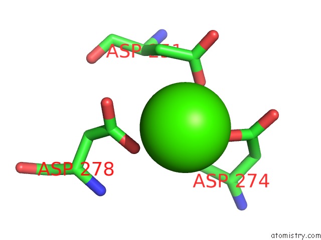
Mono view
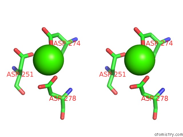
Stereo pair view
Mono view
Stereo pair view
A full contact list of Calcium with other atoms in the Ca binding
site number 2 of Crystal Structure of Trypanosoma Brucei Glycosomal Isocitrate Dehydrogenase in Complex with Nadh, Alpha-Ketoglutarate and CA2+ within 5.0Å range:
|
Calcium binding site 3 out of 4 in 6ajb
Go back to
Calcium binding site 3 out
of 4 in the Crystal Structure of Trypanosoma Brucei Glycosomal Isocitrate Dehydrogenase in Complex with Nadh, Alpha-Ketoglutarate and CA2+
Mono view
Stereo pair view
Mono view
Stereo pair view
A full contact list of Calcium with other atoms in the Ca binding
site number 3 of Crystal Structure of Trypanosoma Brucei Glycosomal Isocitrate Dehydrogenase in Complex with Nadh, Alpha-Ketoglutarate and CA2+ within 5.0Å range:
|
Calcium binding site 4 out of 4 in 6ajb
Go back to
Calcium binding site 4 out
of 4 in the Crystal Structure of Trypanosoma Brucei Glycosomal Isocitrate Dehydrogenase in Complex with Nadh, Alpha-Ketoglutarate and CA2+
Mono view
Stereo pair view
Mono view
Stereo pair view
A full contact list of Calcium with other atoms in the Ca binding
site number 4 of Crystal Structure of Trypanosoma Brucei Glycosomal Isocitrate Dehydrogenase in Complex with Nadh, Alpha-Ketoglutarate and CA2+ within 5.0Å range:
|
Reference:
X.Wang,
D.K.Inaoka,
T.Shiba,
E.O.Balogun,
N.Ziebart,
S.Allman,
Y.Watanabe,
T.Nozaki,
M.Boshart,
F.Bringaud,
S.Harada,
K.Kita.
Biochemical Characterization of A Novel Trypanosoma Brucei Glycosomal Isocitrate Dehydrogenase with Dual Coenzyme Specificity (Nadp+/Nad+) To Be Published.
Page generated: Mon Jul 15 16:29:13 2024
Last articles
Zn in 9MJ5Zn in 9HNW
Zn in 9G0L
Zn in 9FNE
Zn in 9DZN
Zn in 9E0I
Zn in 9D32
Zn in 9DAK
Zn in 8ZXC
Zn in 8ZUF