Calcium »
PDB 6ai0-6b40 »
6amf »
Calcium in PDB 6amf: Crystal Structure of Staphylococcal Nuclease Variant Delta+Phs V23K/L36E at Cryogenic Temperature
Enzymatic activity of Crystal Structure of Staphylococcal Nuclease Variant Delta+Phs V23K/L36E at Cryogenic Temperature
All present enzymatic activity of Crystal Structure of Staphylococcal Nuclease Variant Delta+Phs V23K/L36E at Cryogenic Temperature:
3.1.31.1;
3.1.31.1;
Protein crystallography data
The structure of Crystal Structure of Staphylococcal Nuclease Variant Delta+Phs V23K/L36E at Cryogenic Temperature, PDB code: 6amf
was solved by
A.C.Robinson,
J.L.Schlessman,
B.Garcia-Moreno E.,
with X-Ray Crystallography technique. A brief refinement statistics is given in the table below:
| Resolution Low / High (Å) | 38.46 / 1.85 |
| Space group | P 1 21 1 |
| Cell size a, b, c (Å), α, β, γ (°) | 31.140, 60.316, 38.567, 90.00, 94.32, 90.00 |
| R / Rfree (%) | 20.9 / 26.1 |
Calcium Binding Sites:
The binding sites of Calcium atom in the Crystal Structure of Staphylococcal Nuclease Variant Delta+Phs V23K/L36E at Cryogenic Temperature
(pdb code 6amf). This binding sites where shown within
5.0 Angstroms radius around Calcium atom.
In total only one binding site of Calcium was determined in the Crystal Structure of Staphylococcal Nuclease Variant Delta+Phs V23K/L36E at Cryogenic Temperature, PDB code: 6amf:
In total only one binding site of Calcium was determined in the Crystal Structure of Staphylococcal Nuclease Variant Delta+Phs V23K/L36E at Cryogenic Temperature, PDB code: 6amf:
Calcium binding site 1 out of 1 in 6amf
Go back to
Calcium binding site 1 out
of 1 in the Crystal Structure of Staphylococcal Nuclease Variant Delta+Phs V23K/L36E at Cryogenic Temperature
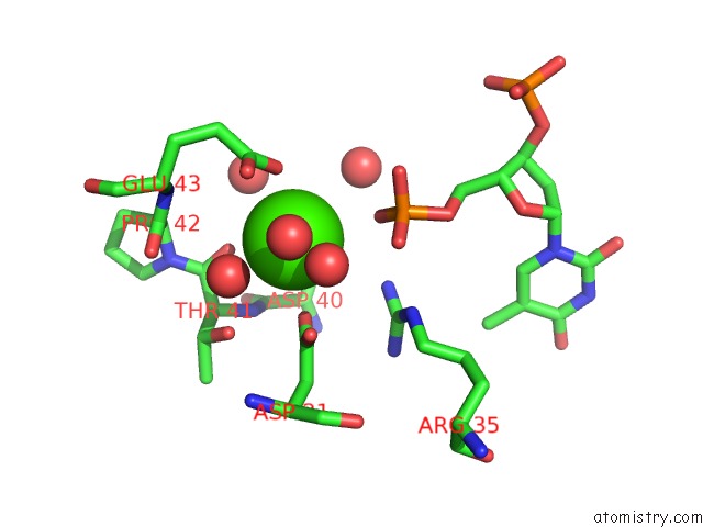
Mono view
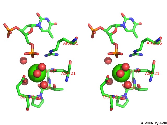
Stereo pair view
Mono view
Stereo pair view
A full contact list of Calcium with other atoms in the Ca binding
site number 1 of Crystal Structure of Staphylococcal Nuclease Variant Delta+Phs V23K/L36E at Cryogenic Temperature within 5.0Å range:
|
Reference:
A.C.Robinson,
J.L.Schlessman,
B.Garcia-Moreno E.
Dielectric Properties of A Protein Probed By Reversal of A Buried Ion Pair. J Phys Chem B V. 122 2516 2018.
ISSN: ISSN 1520-5207
PubMed: 29466010
DOI: 10.1021/ACS.JPCB.7B12121
Page generated: Wed Jul 9 12:31:13 2025
ISSN: ISSN 1520-5207
PubMed: 29466010
DOI: 10.1021/ACS.JPCB.7B12121
Last articles
Ca in 7LG1Ca in 7LCU
Ca in 7LFX
Ca in 7LFW
Ca in 7L8P
Ca in 7LDQ
Ca in 7LAN
Ca in 7LCP
Ca in 7LAG
Ca in 7LAL