Calcium »
PDB 6ai0-6b40 »
6amz »
Calcium in PDB 6amz: Crystal Structure of A Domain Swapped 2,3,4,5-Tetrahydropyridine-2,6- Dicarboxylate N-Succinyltransferase From Acinetobacter Baumannii
Enzymatic activity of Crystal Structure of A Domain Swapped 2,3,4,5-Tetrahydropyridine-2,6- Dicarboxylate N-Succinyltransferase From Acinetobacter Baumannii
All present enzymatic activity of Crystal Structure of A Domain Swapped 2,3,4,5-Tetrahydropyridine-2,6- Dicarboxylate N-Succinyltransferase From Acinetobacter Baumannii:
2.3.1.117;
2.3.1.117;
Protein crystallography data
The structure of Crystal Structure of A Domain Swapped 2,3,4,5-Tetrahydropyridine-2,6- Dicarboxylate N-Succinyltransferase From Acinetobacter Baumannii, PDB code: 6amz
was solved by
T.E.Edwards,
Seattle Structural Genomics Center For Infectious Disease(Ssgcid),
with X-Ray Crystallography technique. A brief refinement statistics is given in the table below:
| Resolution Low / High (Å) | 38.33 / 2.05 |
| Space group | H 3 2 |
| Cell size a, b, c (Å), α, β, γ (°) | 92.250, 92.250, 218.440, 90.00, 90.00, 120.00 |
| R / Rfree (%) | 15.5 / 18.2 |
Calcium Binding Sites:
The binding sites of Calcium atom in the Crystal Structure of A Domain Swapped 2,3,4,5-Tetrahydropyridine-2,6- Dicarboxylate N-Succinyltransferase From Acinetobacter Baumannii
(pdb code 6amz). This binding sites where shown within
5.0 Angstroms radius around Calcium atom.
In total only one binding site of Calcium was determined in the Crystal Structure of A Domain Swapped 2,3,4,5-Tetrahydropyridine-2,6- Dicarboxylate N-Succinyltransferase From Acinetobacter Baumannii, PDB code: 6amz:
In total only one binding site of Calcium was determined in the Crystal Structure of A Domain Swapped 2,3,4,5-Tetrahydropyridine-2,6- Dicarboxylate N-Succinyltransferase From Acinetobacter Baumannii, PDB code: 6amz:
Calcium binding site 1 out of 1 in 6amz
Go back to
Calcium binding site 1 out
of 1 in the Crystal Structure of A Domain Swapped 2,3,4,5-Tetrahydropyridine-2,6- Dicarboxylate N-Succinyltransferase From Acinetobacter Baumannii
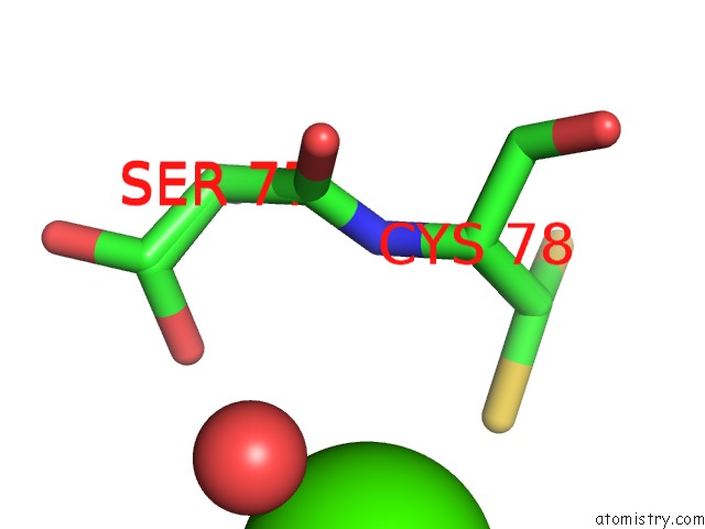
Mono view
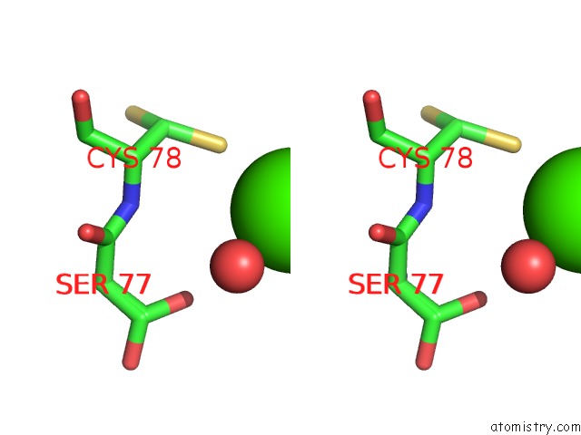
Stereo pair view
Mono view
Stereo pair view
A full contact list of Calcium with other atoms in the Ca binding
site number 1 of Crystal Structure of A Domain Swapped 2,3,4,5-Tetrahydropyridine-2,6- Dicarboxylate N-Succinyltransferase From Acinetobacter Baumannii within 5.0Å range:
|
Reference:
T.E.Edwards,
P.S.Horanyi,
D.D.Lorimer,
Seattle Structural Genomics Center For Infectious Disease.
Crystal Structure of A Domain Swapped 2,3,4,5-Tetrahydropyridine-2,6-Dicarboxylate N-Succinyltransferase From Acinetobacter Baumannii To Be Published.
Page generated: Wed Jul 9 12:31:27 2025
Last articles
F in 4JKVF in 4JLJ
F in 4JJS
F in 4JII
F in 4JDS
F in 4JAV
F in 4JA8
F in 4JBY
F in 4JB4
F in 4JAS