Calcium »
PDB 6b5v-6bia »
6bia »
Calcium in PDB 6bia: Crystal Structure of Ps I-Cgsb
Protein crystallography data
The structure of Crystal Structure of Ps I-Cgsb, PDB code: 6bia
was solved by
A.G.Hettle,
A.B.Boraston,
with X-Ray Crystallography technique. A brief refinement statistics is given in the table below:
| Resolution Low / High (Å) | 114.86 / 2.80 |
| Space group | P 43 21 2 |
| Cell size a, b, c (Å), α, β, γ (°) | 133.850, 133.850, 223.695, 90.00, 90.00, 90.00 |
| R / Rfree (%) | 23.8 / 26.5 |
Calcium Binding Sites:
The binding sites of Calcium atom in the Crystal Structure of Ps I-Cgsb
(pdb code 6bia). This binding sites where shown within
5.0 Angstroms radius around Calcium atom.
In total 3 binding sites of Calcium where determined in the Crystal Structure of Ps I-Cgsb, PDB code: 6bia:
Jump to Calcium binding site number: 1; 2; 3;
In total 3 binding sites of Calcium where determined in the Crystal Structure of Ps I-Cgsb, PDB code: 6bia:
Jump to Calcium binding site number: 1; 2; 3;
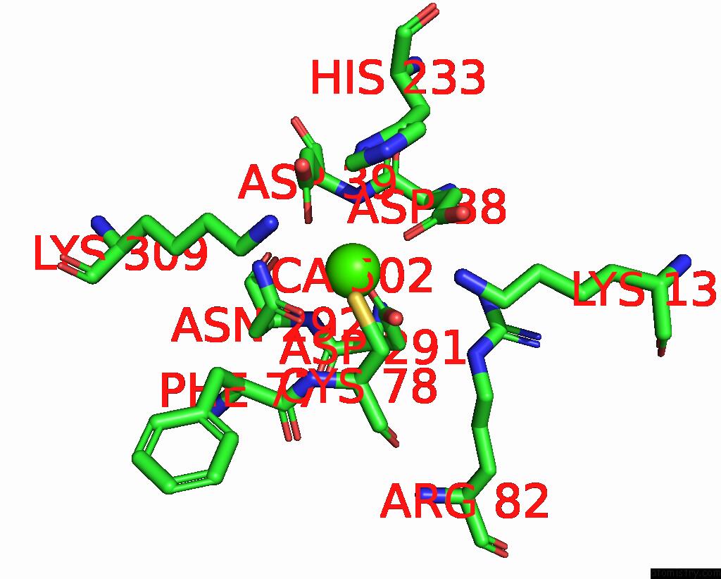
Calcium binding site 1 out of 3 in 6bia
Go back to
Calcium binding site 1 out
of 3 in the Crystal Structure of Ps I-Cgsb
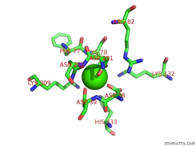
Mono view
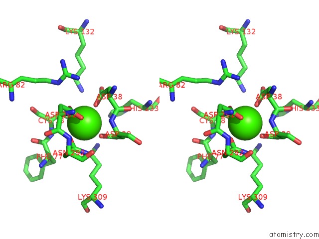
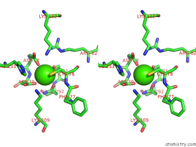
Stereo pair view
Mono view
Stereo pair view
A full contact list of Calcium with other atoms in the Ca binding
site number 1 of Crystal Structure of Ps I-Cgsb within 5.0Å range:
|
Calcium binding site 2 out of 3 in 6bia
Go back to
Calcium binding site 2 out
of 3 in the Crystal Structure of Ps I-Cgsb
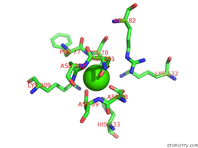
Mono view
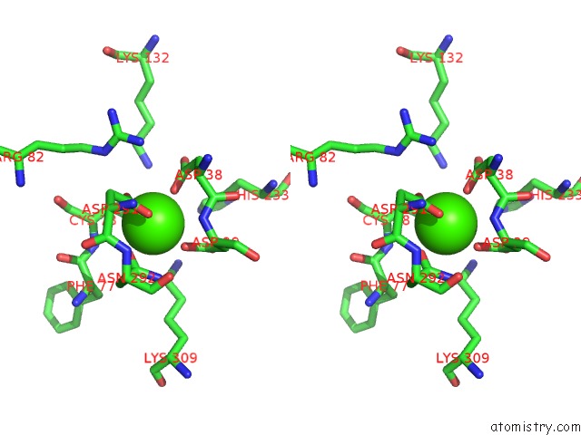
Stereo pair view
Mono view
Stereo pair view
A full contact list of Calcium with other atoms in the Ca binding
site number 2 of Crystal Structure of Ps I-Cgsb within 5.0Å range:
|
Calcium binding site 3 out of 3 in 6bia
Go back to
Calcium binding site 3 out
of 3 in the Crystal Structure of Ps I-Cgsb
Mono view
Stereo pair view
Mono view
Stereo pair view
A full contact list of Calcium with other atoms in the Ca binding
site number 3 of Crystal Structure of Ps I-Cgsb within 5.0Å range:
|
Reference:
A.G.Hettle,
C.Vickers,
C.S.Robb,
F.Liu,
S.G.Withers,
J.H.Hehemann,
A.B.Boraston.
The Molecular Basis of Polysaccharide Sulfatase Activity and A Nomenclature For Catalytic Subsites in This Class of Enzyme. Structure V. 26 747 2018.
ISSN: ISSN 1878-4186
PubMed: 29681469
DOI: 10.1016/J.STR.2018.03.012
Page generated: Wed Jul 9 12:48:16 2025
ISSN: ISSN 1878-4186
PubMed: 29681469
DOI: 10.1016/J.STR.2018.03.012
Last articles
Cl in 5L79Cl in 5L77
Cl in 5L6J
Cl in 5L74
Cl in 5L6Q
Cl in 5L6C
Cl in 5L69
Cl in 5L6B
Cl in 5L6A
Cl in 5L66