Calcium »
PDB 6bjd-6bxz »
6bur »
Calcium in PDB 6bur: Crystal Structures of Cyanuric Acid Hydrolase From Moorella Thermoacetica Complexed with Barbituric Acid
Enzymatic activity of Crystal Structures of Cyanuric Acid Hydrolase From Moorella Thermoacetica Complexed with Barbituric Acid
All present enzymatic activity of Crystal Structures of Cyanuric Acid Hydrolase From Moorella Thermoacetica Complexed with Barbituric Acid:
3.5.2.15;
3.5.2.15;
Protein crystallography data
The structure of Crystal Structures of Cyanuric Acid Hydrolase From Moorella Thermoacetica Complexed with Barbituric Acid, PDB code: 6bur
was solved by
K.Shi,
H.Aihara,
with X-Ray Crystallography technique. A brief refinement statistics is given in the table below:
| Resolution Low / High (Å) | 39.72 / 2.18 |
| Space group | P 21 21 21 |
| Cell size a, b, c (Å), α, β, γ (°) | 81.075, 89.121, 199.121, 90.00, 90.00, 90.00 |
| R / Rfree (%) | 16.3 / 21.2 |
Calcium Binding Sites:
The binding sites of Calcium atom in the Crystal Structures of Cyanuric Acid Hydrolase From Moorella Thermoacetica Complexed with Barbituric Acid
(pdb code 6bur). This binding sites where shown within
5.0 Angstroms radius around Calcium atom.
In total 4 binding sites of Calcium where determined in the Crystal Structures of Cyanuric Acid Hydrolase From Moorella Thermoacetica Complexed with Barbituric Acid, PDB code: 6bur:
Jump to Calcium binding site number: 1; 2; 3; 4;
In total 4 binding sites of Calcium where determined in the Crystal Structures of Cyanuric Acid Hydrolase From Moorella Thermoacetica Complexed with Barbituric Acid, PDB code: 6bur:
Jump to Calcium binding site number: 1; 2; 3; 4;
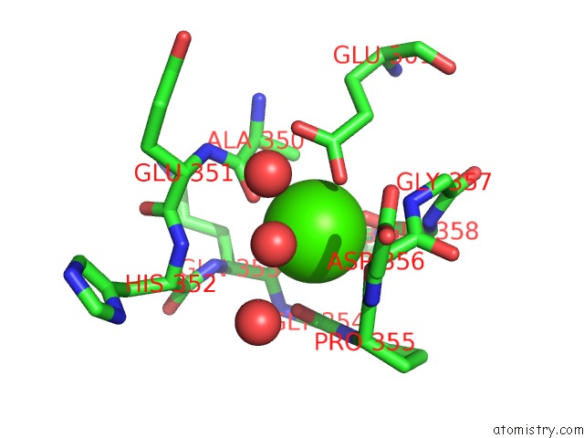
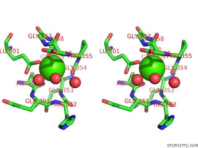
Calcium binding site 1 out of 4 in 6bur
Go back to
Calcium binding site 1 out
of 4 in the Crystal Structures of Cyanuric Acid Hydrolase From Moorella Thermoacetica Complexed with Barbituric Acid
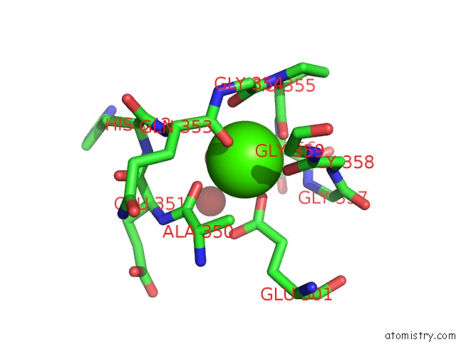
Mono view
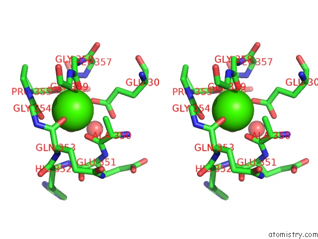
Stereo pair view
Mono view
Stereo pair view
A full contact list of Calcium with other atoms in the Ca binding
site number 1 of Crystal Structures of Cyanuric Acid Hydrolase From Moorella Thermoacetica Complexed with Barbituric Acid within 5.0Å range:
|
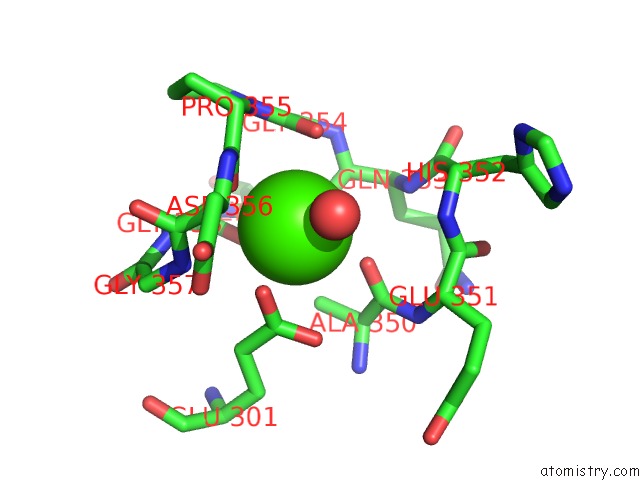
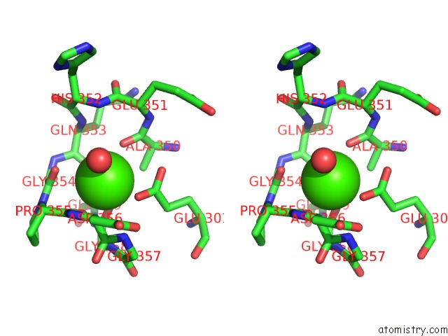
Calcium binding site 2 out of 4 in 6bur
Go back to
Calcium binding site 2 out
of 4 in the Crystal Structures of Cyanuric Acid Hydrolase From Moorella Thermoacetica Complexed with Barbituric Acid
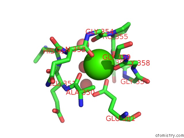
Mono view
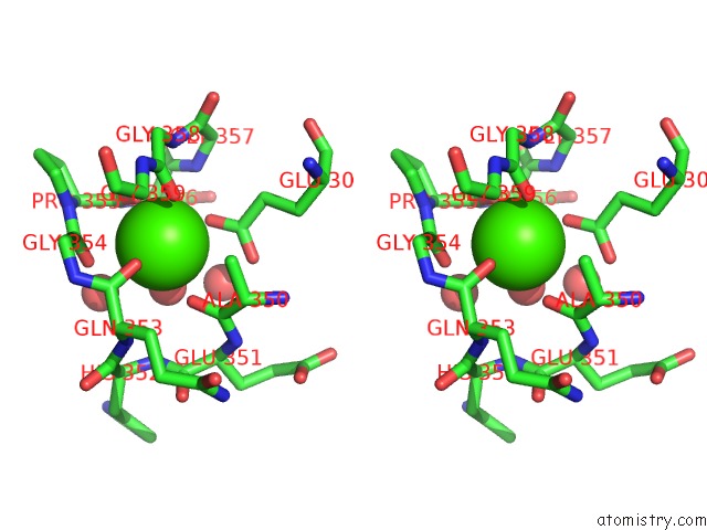
Stereo pair view
Mono view
Stereo pair view
A full contact list of Calcium with other atoms in the Ca binding
site number 2 of Crystal Structures of Cyanuric Acid Hydrolase From Moorella Thermoacetica Complexed with Barbituric Acid within 5.0Å range:
|
Calcium binding site 3 out of 4 in 6bur
Go back to
Calcium binding site 3 out
of 4 in the Crystal Structures of Cyanuric Acid Hydrolase From Moorella Thermoacetica Complexed with Barbituric Acid
Mono view
Stereo pair view
Mono view
Stereo pair view
A full contact list of Calcium with other atoms in the Ca binding
site number 3 of Crystal Structures of Cyanuric Acid Hydrolase From Moorella Thermoacetica Complexed with Barbituric Acid within 5.0Å range:
|
Calcium binding site 4 out of 4 in 6bur
Go back to
Calcium binding site 4 out
of 4 in the Crystal Structures of Cyanuric Acid Hydrolase From Moorella Thermoacetica Complexed with Barbituric Acid
Mono view
Stereo pair view
Mono view
Stereo pair view
A full contact list of Calcium with other atoms in the Ca binding
site number 4 of Crystal Structures of Cyanuric Acid Hydrolase From Moorella Thermoacetica Complexed with Barbituric Acid within 5.0Å range:
|
Reference:
K.Shi,
S.Cho,
B.Asim,
J.L.Seffernick,
L.P.Wackett,
H.Aihara.
Crystal Structures of Cyanuric Acid Hydrolase From Moorella Thermoacetica To Be Published.
Page generated: Wed Jul 9 12:55:24 2025
Last articles
F in 7NVOF in 7NTH
F in 7NTI
F in 7NPC
F in 7NRG
F in 7NR5
F in 7NQS
F in 7NOS
F in 7NP5
F in 7NDV