Calcium »
PDB 6bjd-6bxz »
6bvd »
Calcium in PDB 6bvd: Structure of Botulinum Neurotoxin Serotype Ha Light Chain
Protein crystallography data
The structure of Structure of Botulinum Neurotoxin Serotype Ha Light Chain, PDB code: 6bvd
was solved by
R.Jin,
K.Lam,
with X-Ray Crystallography technique. A brief refinement statistics is given in the table below:
| Resolution Low / High (Å) | 54.05 / 2.09 |
| Space group | C 2 2 21 |
| Cell size a, b, c (Å), α, β, γ (°) | 76.249, 160.745, 219.099, 90.00, 90.00, 90.00 |
| R / Rfree (%) | 17.6 / 19.1 |
Other elements in 6bvd:
The structure of Structure of Botulinum Neurotoxin Serotype Ha Light Chain also contains other interesting chemical elements:
| Zinc | (Zn) | 2 atoms |
Calcium Binding Sites:
The binding sites of Calcium atom in the Structure of Botulinum Neurotoxin Serotype Ha Light Chain
(pdb code 6bvd). This binding sites where shown within
5.0 Angstroms radius around Calcium atom.
In total only one binding site of Calcium was determined in the Structure of Botulinum Neurotoxin Serotype Ha Light Chain, PDB code: 6bvd:
In total only one binding site of Calcium was determined in the Structure of Botulinum Neurotoxin Serotype Ha Light Chain, PDB code: 6bvd:
Calcium binding site 1 out of 1 in 6bvd
Go back to
Calcium binding site 1 out
of 1 in the Structure of Botulinum Neurotoxin Serotype Ha Light Chain
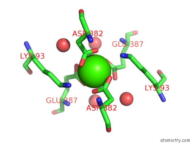
Mono view
Stereo pair view
Mono view
Stereo pair view
A full contact list of Calcium with other atoms in the Ca binding
site number 1 of Structure of Botulinum Neurotoxin Serotype Ha Light Chain within 5.0Å range:
|
Reference:
K.H.Lam,
S.Sikorra,
J.Weisemann,
H.Maatsch,
K.Perry,
A.Rummel,
T.Binz,
R.Jin.
Structural and Biochemical Characterization of the Protease Domain of the Mosaic Botulinum Neurotoxin Type Ha. Pathog Dis V. 76 2018.
ISSN: ISSN 2049-632X
PubMed: 29688327
DOI: 10.1093/FEMSPD/FTY044
Page generated: Mon Jul 15 17:10:11 2024
ISSN: ISSN 2049-632X
PubMed: 29688327
DOI: 10.1093/FEMSPD/FTY044
Last articles
Zn in 9MJ5Zn in 9HNW
Zn in 9G0L
Zn in 9FNE
Zn in 9DZN
Zn in 9E0I
Zn in 9D32
Zn in 9DAK
Zn in 8ZXC
Zn in 8ZUF