Calcium »
PDB 6bjd-6bxz »
6bxu »
Calcium in PDB 6bxu: Crystal Structure of Mouse Protocadherin-15 EC5-7 I582T
Protein crystallography data
The structure of Crystal Structure of Mouse Protocadherin-15 EC5-7 I582T, PDB code: 6bxu
was solved by
B.L.Neel,
M.Sotomayor,
with X-Ray Crystallography technique. A brief refinement statistics is given in the table below:
| Resolution Low / High (Å) | 180.90 / 3.79 |
| Space group | P 42 2 2 |
| Cell size a, b, c (Å), α, β, γ (°) | 180.905, 180.905, 127.161, 90.00, 90.00, 90.00 |
| R / Rfree (%) | 24 / 28.3 |
Calcium Binding Sites:
The binding sites of Calcium atom in the Crystal Structure of Mouse Protocadherin-15 EC5-7 I582T
(pdb code 6bxu). This binding sites where shown within
5.0 Angstroms radius around Calcium atom.
In total 10 binding sites of Calcium where determined in the Crystal Structure of Mouse Protocadherin-15 EC5-7 I582T, PDB code: 6bxu:
Jump to Calcium binding site number: 1; 2; 3; 4; 5; 6; 7; 8; 9; 10;
In total 10 binding sites of Calcium where determined in the Crystal Structure of Mouse Protocadherin-15 EC5-7 I582T, PDB code: 6bxu:
Jump to Calcium binding site number: 1; 2; 3; 4; 5; 6; 7; 8; 9; 10;
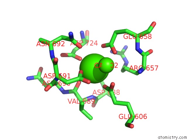
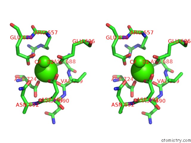
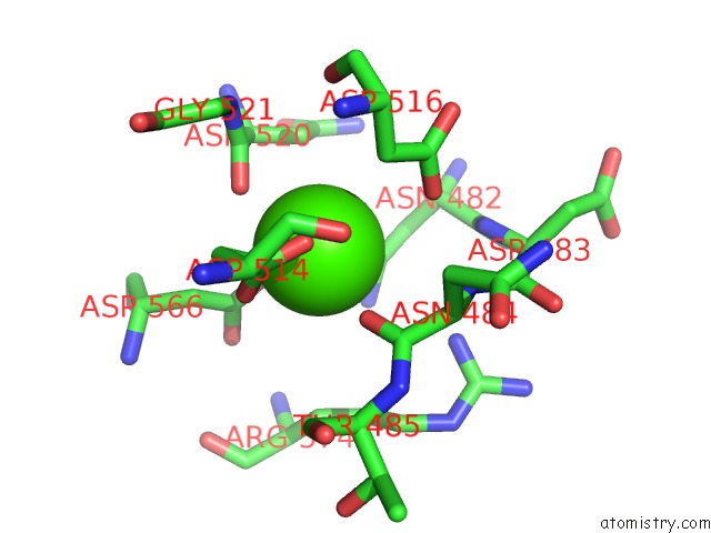
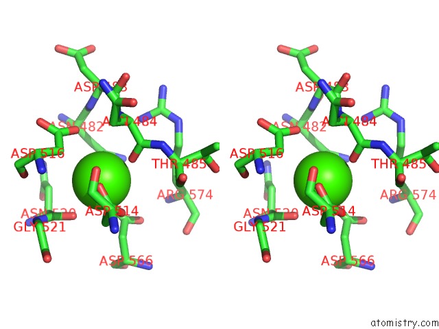
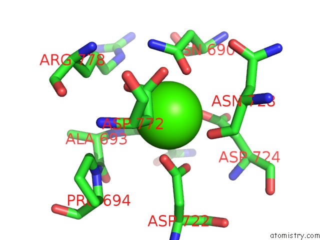
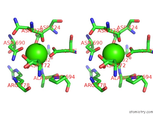
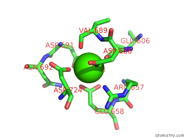
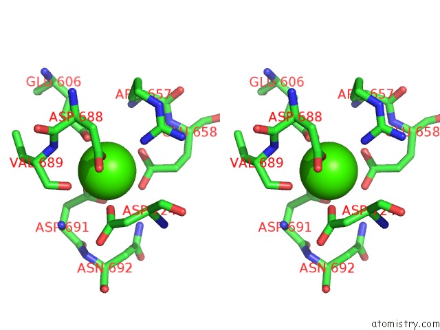
Calcium binding site 1 out of 10 in 6bxu
Go back to
Calcium binding site 1 out
of 10 in the Crystal Structure of Mouse Protocadherin-15 EC5-7 I582T
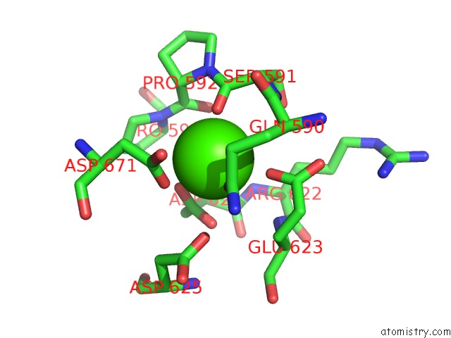
Mono view
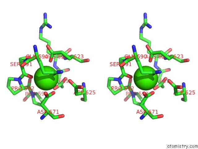
Stereo pair view
Mono view
Stereo pair view
|
|
A full contact list of Calcium with other atoms in the Ca binding
site number 1 of Crystal Structure of Mouse Protocadherin-15 EC5-7 I582T within 5.0Å range:
|
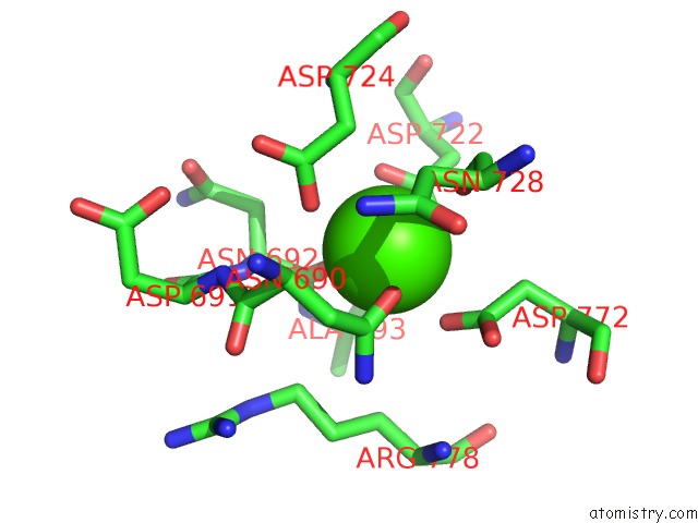
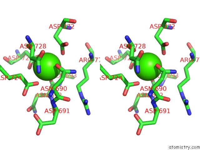
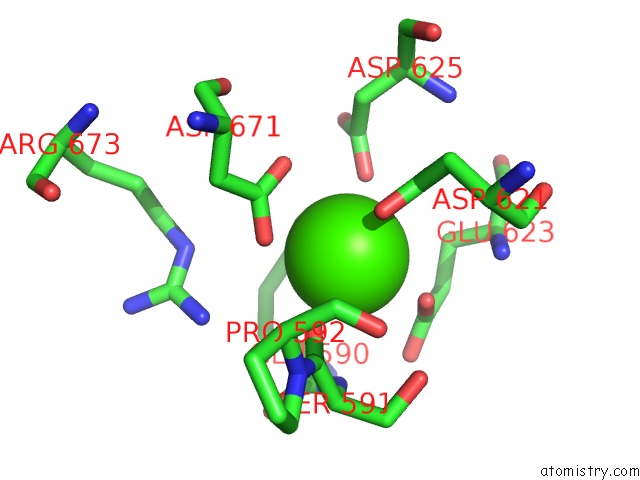
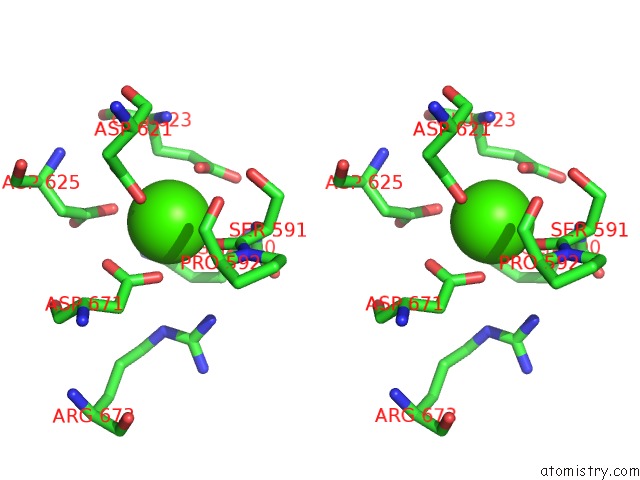
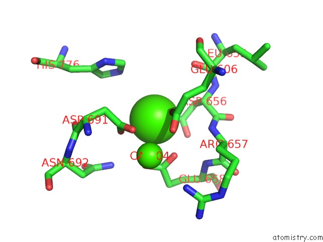
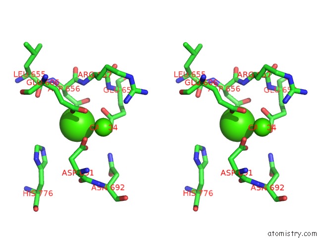
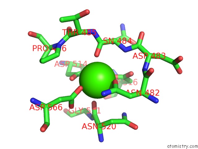
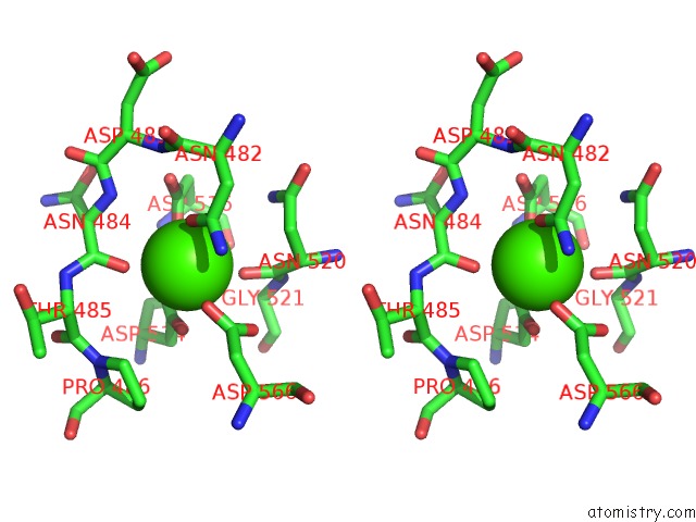
Calcium binding site 2 out of 10 in 6bxu
Go back to
Calcium binding site 2 out
of 10 in the Crystal Structure of Mouse Protocadherin-15 EC5-7 I582T
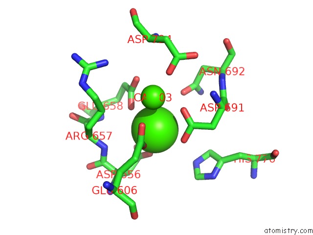
Mono view
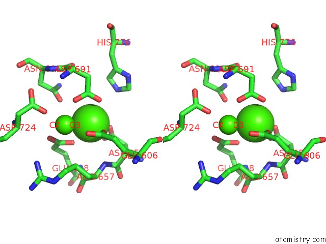
Stereo pair view
Mono view
Stereo pair view
|
|
A full contact list of Calcium with other atoms in the Ca binding
site number 2 of Crystal Structure of Mouse Protocadherin-15 EC5-7 I582T within 5.0Å range:
|
Calcium binding site 3 out of 10 in 6bxu
Go back to
Calcium binding site 3 out
of 10 in the Crystal Structure of Mouse Protocadherin-15 EC5-7 I582T
Mono view
Stereo pair view
Mono view
Stereo pair view
|
|
A full contact list of Calcium with other atoms in the Ca binding
site number 3 of Crystal Structure of Mouse Protocadherin-15 EC5-7 I582T within 5.0Å range:
|
Calcium binding site 4 out of 10 in 6bxu
Go back to
Calcium binding site 4 out
of 10 in the Crystal Structure of Mouse Protocadherin-15 EC5-7 I582T
Mono view
Stereo pair view
Mono view
Stereo pair view
|
|
A full contact list of Calcium with other atoms in the Ca binding
site number 4 of Crystal Structure of Mouse Protocadherin-15 EC5-7 I582T within 5.0Å range:
|
Calcium binding site 5 out of 10 in 6bxu
Go back to
Calcium binding site 5 out
of 10 in the Crystal Structure of Mouse Protocadherin-15 EC5-7 I582T
Mono view
Stereo pair view
Mono view
Stereo pair view
|
|
A full contact list of Calcium with other atoms in the Ca binding
site number 5 of Crystal Structure of Mouse Protocadherin-15 EC5-7 I582T within 5.0Å range:
|
Calcium binding site 6 out of 10 in 6bxu
Go back to
Calcium binding site 6 out
of 10 in the Crystal Structure of Mouse Protocadherin-15 EC5-7 I582T
Mono view
Stereo pair view
Mono view
Stereo pair view
|
|
A full contact list of Calcium with other atoms in the Ca binding
site number 6 of Crystal Structure of Mouse Protocadherin-15 EC5-7 I582T within 5.0Å range:
|
Calcium binding site 7 out of 10 in 6bxu
Go back to
Calcium binding site 7 out
of 10 in the Crystal Structure of Mouse Protocadherin-15 EC5-7 I582T
Mono view
Stereo pair view
Mono view
Stereo pair view
|
|
A full contact list of Calcium with other atoms in the Ca binding
site number 7 of Crystal Structure of Mouse Protocadherin-15 EC5-7 I582T within 5.0Å range:
|
Calcium binding site 8 out of 10 in 6bxu
Go back to
Calcium binding site 8 out
of 10 in the Crystal Structure of Mouse Protocadherin-15 EC5-7 I582T
Mono view
Stereo pair view
Mono view
Stereo pair view
|
|
A full contact list of Calcium with other atoms in the Ca binding
site number 8 of Crystal Structure of Mouse Protocadherin-15 EC5-7 I582T within 5.0Å range:
|
Calcium binding site 9 out of 10 in 6bxu
Go back to
Calcium binding site 9 out
of 10 in the Crystal Structure of Mouse Protocadherin-15 EC5-7 I582T
Mono view
Stereo pair view
Mono view
Stereo pair view
|
|
A full contact list of Calcium with other atoms in the Ca binding
site number 9 of Crystal Structure of Mouse Protocadherin-15 EC5-7 I582T within 5.0Å range:
|
Calcium binding site 10 out of 10 in 6bxu
Go back to
Calcium binding site 10 out
of 10 in the Crystal Structure of Mouse Protocadherin-15 EC5-7 I582T
Mono view
Stereo pair view
Mono view
Stereo pair view
|
|
A full contact list of Calcium with other atoms in the Ca binding
site number 10 of Crystal Structure of Mouse Protocadherin-15 EC5-7 I582T within 5.0Å range:
|
Reference:
B.L.Neel,
M.Sotomayor.
Crystal Structure of Mouse Protocadherin-15 EC5-7 I582T To Be Published.
Page generated: Mon Jul 15 17:12:30 2024
Last articles
Zn in 9J0NZn in 9J0O
Zn in 9J0P
Zn in 9FJX
Zn in 9EKB
Zn in 9C0F
Zn in 9CAH
Zn in 9CH0
Zn in 9CH3
Zn in 9CH1