Calcium »
PDB 6byp-6cl6 »
6c2h »
Calcium in PDB 6c2h: Crystal Structures of Cystathionine Beta-Synthase From Saccharomyces Cerevisiae: the Structure of the Catalytic Core
Enzymatic activity of Crystal Structures of Cystathionine Beta-Synthase From Saccharomyces Cerevisiae: the Structure of the Catalytic Core
All present enzymatic activity of Crystal Structures of Cystathionine Beta-Synthase From Saccharomyces Cerevisiae: the Structure of the Catalytic Core:
4.2.1.22;
4.2.1.22;
Protein crystallography data
The structure of Crystal Structures of Cystathionine Beta-Synthase From Saccharomyces Cerevisiae: the Structure of the Catalytic Core, PDB code: 6c2h
was solved by
C.A.Kreinbring,
Y.Tu,
D.Liu,
G.A.Petsko,
D.Ringe,
with X-Ray Crystallography technique. A brief refinement statistics is given in the table below:
| Resolution Low / High (Å) | 50.00 / 1.49 |
| Space group | P 65 2 2 |
| Cell size a, b, c (Å), α, β, γ (°) | 81.374, 81.374, 208.440, 90.00, 90.00, 120.00 |
| R / Rfree (%) | 12.6 / 16.3 |
Other elements in 6c2h:
The structure of Crystal Structures of Cystathionine Beta-Synthase From Saccharomyces Cerevisiae: the Structure of the Catalytic Core also contains other interesting chemical elements:
| Chlorine | (Cl) | 1 atom |
| Sodium | (Na) | 1 atom |
Calcium Binding Sites:
The binding sites of Calcium atom in the Crystal Structures of Cystathionine Beta-Synthase From Saccharomyces Cerevisiae: the Structure of the Catalytic Core
(pdb code 6c2h). This binding sites where shown within
5.0 Angstroms radius around Calcium atom.
In total only one binding site of Calcium was determined in the Crystal Structures of Cystathionine Beta-Synthase From Saccharomyces Cerevisiae: the Structure of the Catalytic Core, PDB code: 6c2h:
In total only one binding site of Calcium was determined in the Crystal Structures of Cystathionine Beta-Synthase From Saccharomyces Cerevisiae: the Structure of the Catalytic Core, PDB code: 6c2h:
Calcium binding site 1 out of 1 in 6c2h
Go back to
Calcium binding site 1 out
of 1 in the Crystal Structures of Cystathionine Beta-Synthase From Saccharomyces Cerevisiae: the Structure of the Catalytic Core
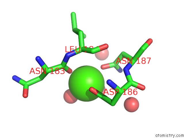
Mono view
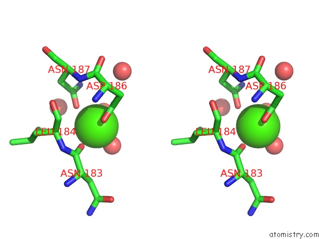
Stereo pair view
Mono view
Stereo pair view
A full contact list of Calcium with other atoms in the Ca binding
site number 1 of Crystal Structures of Cystathionine Beta-Synthase From Saccharomyces Cerevisiae: the Structure of the Catalytic Core within 5.0Å range:
|
Reference:
Y.Tu,
C.A.Kreinbring,
M.Hill,
C.Liu,
G.A.Petsko,
C.D.Mccune,
D.B.Berkowitz,
D.Liu,
D.Ringe.
Crystal Structures of Cystathionine Beta-Synthase From Saccharomyces Cerevisiae: One Enzymatic Step at A Time. Biochemistry V. 57 3134 2018.
ISSN: ISSN 1520-4995
PubMed: 29630349
DOI: 10.1021/ACS.BIOCHEM.8B00092
Page generated: Mon Jul 15 17:19:16 2024
ISSN: ISSN 1520-4995
PubMed: 29630349
DOI: 10.1021/ACS.BIOCHEM.8B00092
Last articles
Zn in 9J0NZn in 9J0O
Zn in 9J0P
Zn in 9FJX
Zn in 9EKB
Zn in 9C0F
Zn in 9CAH
Zn in 9CH0
Zn in 9CH3
Zn in 9CH1