Calcium »
PDB 6bxz-6ckm »
6c74 »
Calcium in PDB 6c74: Crystal Structure of Murine CD300LF in Complex with Phosphocholine
Protein crystallography data
The structure of Crystal Structure of Murine CD300LF in Complex with Phosphocholine, PDB code: 6c74
was solved by
C.A.Nelson,
D.H.Fremont,
with X-Ray Crystallography technique. A brief refinement statistics is given in the table below:
| Resolution Low / High (Å) | 34.55 / 1.36 |
| Space group | P 21 21 21 |
| Cell size a, b, c (Å), α, β, γ (°) | 32.640, 40.058, 68.291, 90.00, 90.00, 90.00 |
| R / Rfree (%) | 16.5 / 18.9 |
Calcium Binding Sites:
The binding sites of Calcium atom in the Crystal Structure of Murine CD300LF in Complex with Phosphocholine
(pdb code 6c74). This binding sites where shown within
5.0 Angstroms radius around Calcium atom.
In total only one binding site of Calcium was determined in the Crystal Structure of Murine CD300LF in Complex with Phosphocholine, PDB code: 6c74:
In total only one binding site of Calcium was determined in the Crystal Structure of Murine CD300LF in Complex with Phosphocholine, PDB code: 6c74:
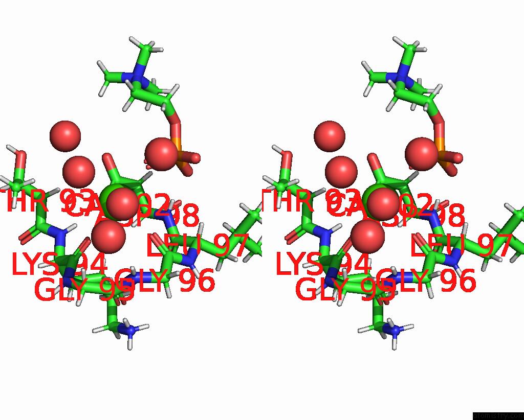
Calcium binding site 1 out of 1 in 6c74
Go back to
Calcium binding site 1 out
of 1 in the Crystal Structure of Murine CD300LF in Complex with Phosphocholine

Mono view
Stereo pair view
Mono view
Stereo pair view
A full contact list of Calcium with other atoms in the Ca binding
site number 1 of Crystal Structure of Murine CD300LF in Complex with Phosphocholine within 5.0Å range:
|
Reference:
C.A.Nelson,
C.B.Wilen,
Y.N.Dai,
R.C.Orchard,
A.S.Kim,
R.A.Stegeman,
L.L.Hsieh,
T.J.Smith,
H.W.Virgin,
D.H.Fremont.
Structural Basis For Murine Norovirus Engagement of Bile Acids and the CD300LF Receptor. Proc. Natl. Acad. Sci. V. 115 E9201 2018U.S.A..
ISSN: ESSN 1091-6490
PubMed: 30194229
DOI: 10.1073/PNAS.1805797115
Page generated: Mon Jul 15 17:20:38 2024
ISSN: ESSN 1091-6490
PubMed: 30194229
DOI: 10.1073/PNAS.1805797115
Last articles
Zn in 9JYWZn in 9IR4
Zn in 9IR3
Zn in 9GMX
Zn in 9GMW
Zn in 9JEJ
Zn in 9ERF
Zn in 9ERE
Zn in 9EGV
Zn in 9EGW