Calcium »
PDB 6dmn-6e48 »
6e1m »
Calcium in PDB 6e1m: Structure of ATTPC1(Dde) Reconstituted in Saposin A
Calcium Binding Sites:
The binding sites of Calcium atom in the Structure of ATTPC1(Dde) Reconstituted in Saposin A
(pdb code 6e1m). This binding sites where shown within
5.0 Angstroms radius around Calcium atom.
In total 7 binding sites of Calcium where determined in the Structure of ATTPC1(Dde) Reconstituted in Saposin A, PDB code: 6e1m:
Jump to Calcium binding site number: 1; 2; 3; 4; 5; 6; 7;
In total 7 binding sites of Calcium where determined in the Structure of ATTPC1(Dde) Reconstituted in Saposin A, PDB code: 6e1m:
Jump to Calcium binding site number: 1; 2; 3; 4; 5; 6; 7;
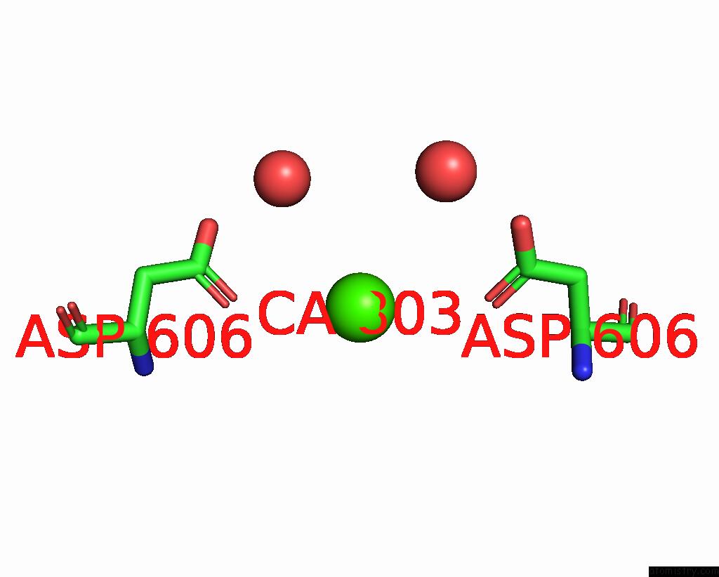
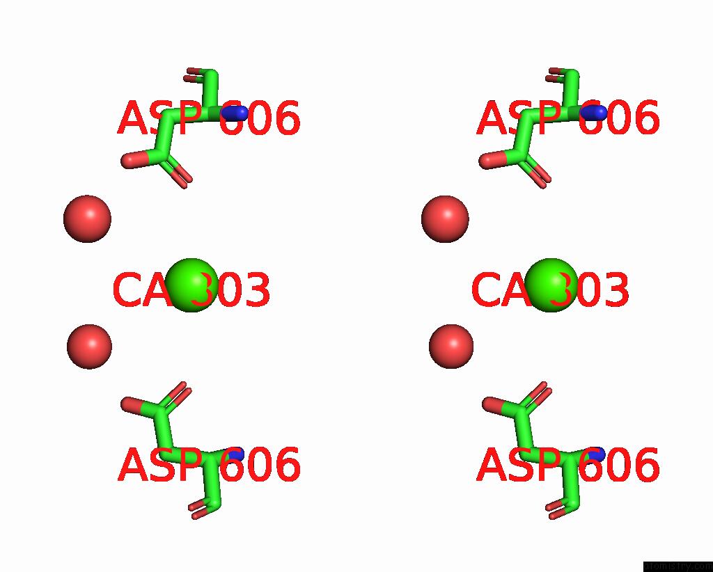
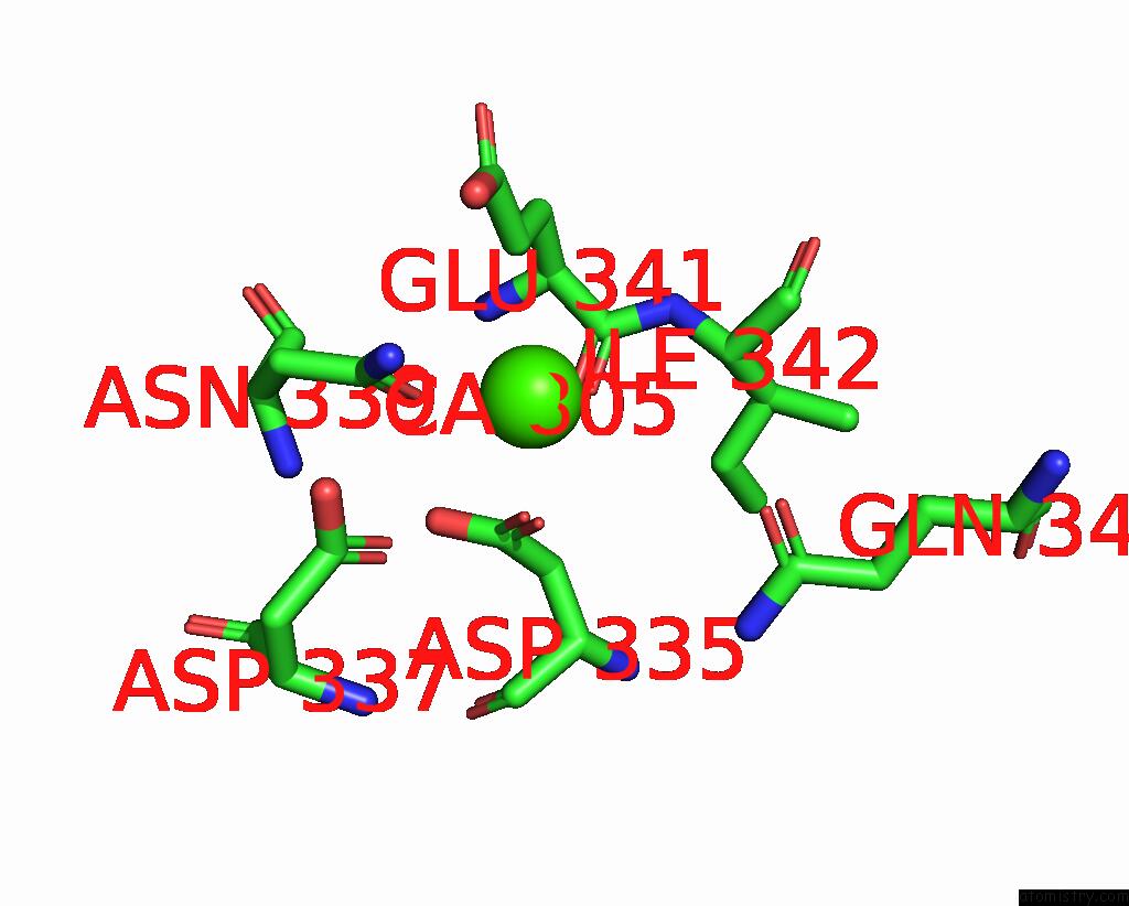
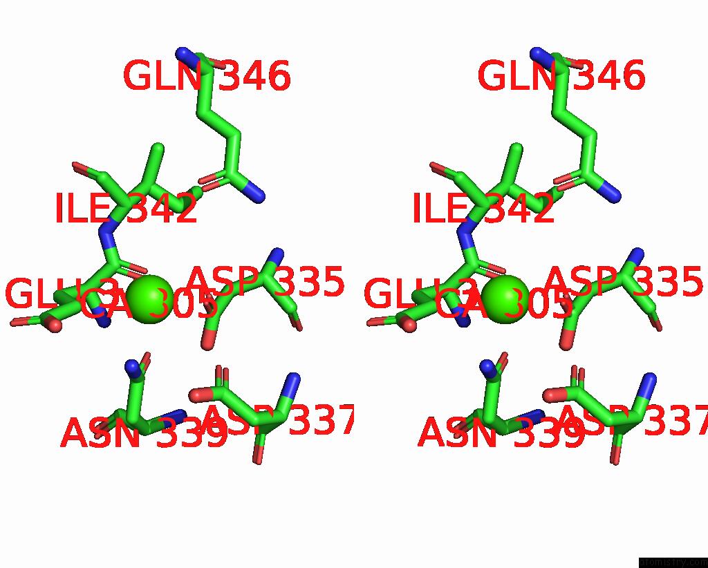
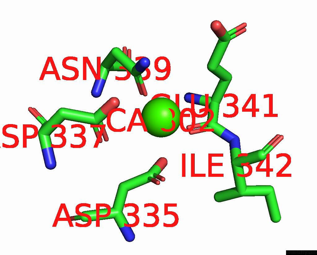
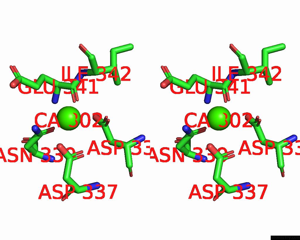
Calcium binding site 1 out of 7 in 6e1m
Go back to
Calcium binding site 1 out
of 7 in the Structure of ATTPC1(Dde) Reconstituted in Saposin A
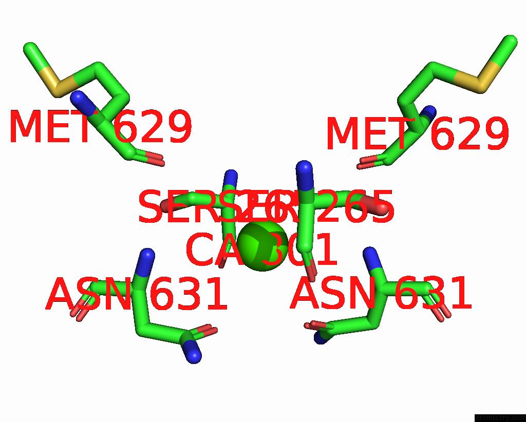
Mono view
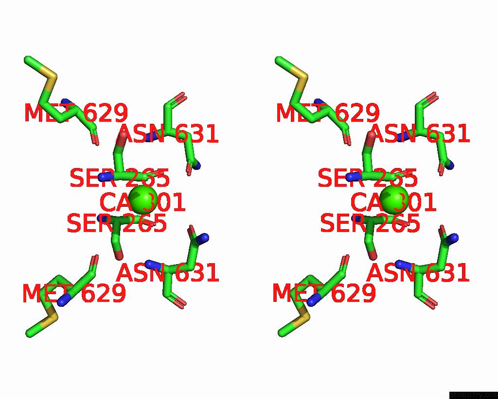
Stereo pair view
Mono view
Stereo pair view
A full contact list of Calcium with other atoms in the Ca binding
site number 1 of Structure of ATTPC1(Dde) Reconstituted in Saposin A within 5.0Å range:
|
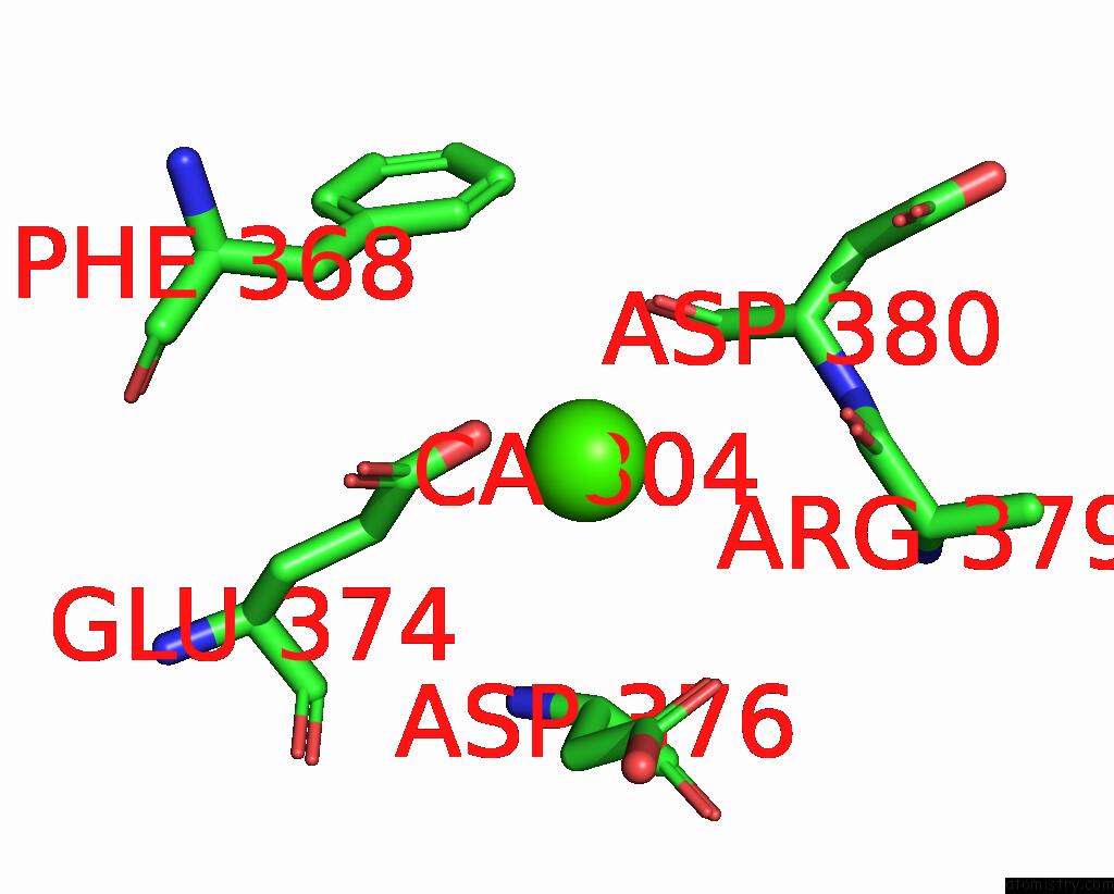
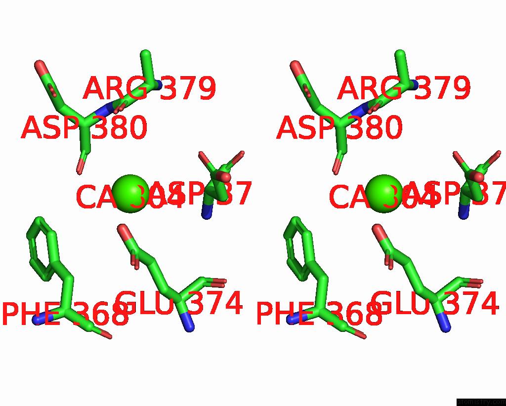
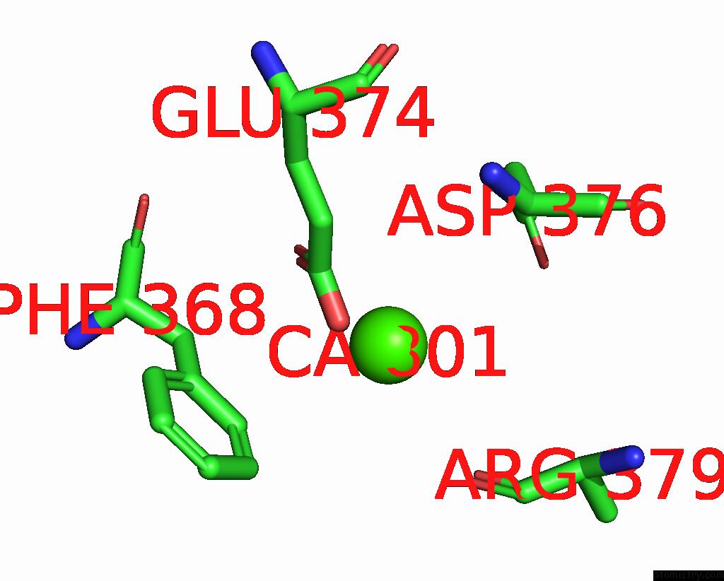
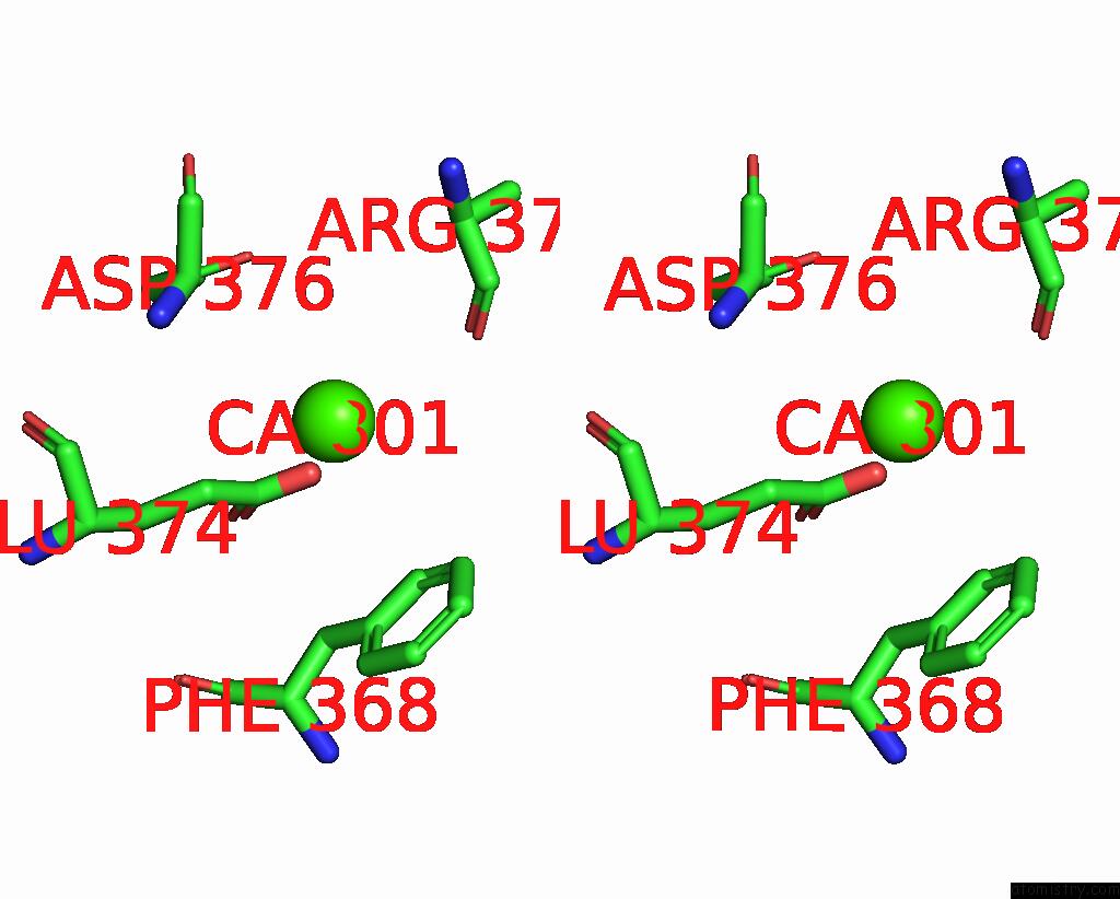
Calcium binding site 2 out of 7 in 6e1m
Go back to
Calcium binding site 2 out
of 7 in the Structure of ATTPC1(Dde) Reconstituted in Saposin A
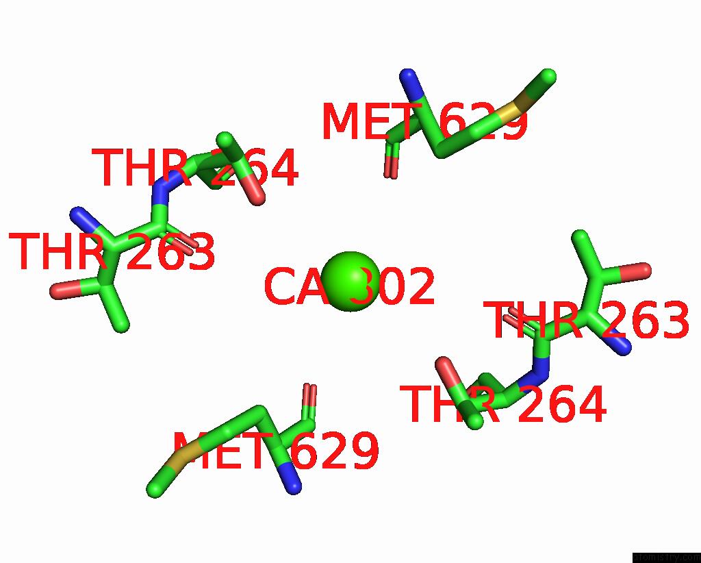
Mono view
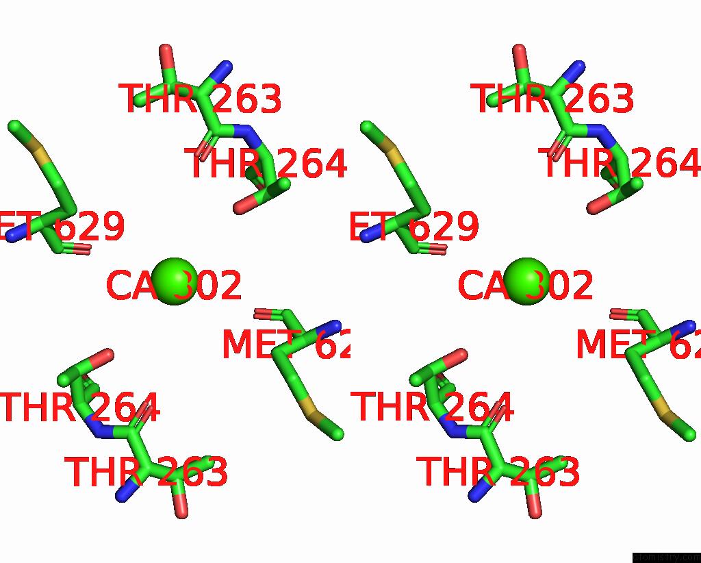
Stereo pair view
Mono view
Stereo pair view
A full contact list of Calcium with other atoms in the Ca binding
site number 2 of Structure of ATTPC1(Dde) Reconstituted in Saposin A within 5.0Å range:
|
Calcium binding site 3 out of 7 in 6e1m
Go back to
Calcium binding site 3 out
of 7 in the Structure of ATTPC1(Dde) Reconstituted in Saposin A
Mono view
Stereo pair view
Mono view
Stereo pair view
A full contact list of Calcium with other atoms in the Ca binding
site number 3 of Structure of ATTPC1(Dde) Reconstituted in Saposin A within 5.0Å range:
|
Calcium binding site 4 out of 7 in 6e1m
Go back to
Calcium binding site 4 out
of 7 in the Structure of ATTPC1(Dde) Reconstituted in Saposin A
Mono view
Stereo pair view
Mono view
Stereo pair view
A full contact list of Calcium with other atoms in the Ca binding
site number 4 of Structure of ATTPC1(Dde) Reconstituted in Saposin A within 5.0Å range:
|
Calcium binding site 5 out of 7 in 6e1m
Go back to
Calcium binding site 5 out
of 7 in the Structure of ATTPC1(Dde) Reconstituted in Saposin A
Mono view
Stereo pair view
Mono view
Stereo pair view
A full contact list of Calcium with other atoms in the Ca binding
site number 5 of Structure of ATTPC1(Dde) Reconstituted in Saposin A within 5.0Å range:
|
Calcium binding site 6 out of 7 in 6e1m
Go back to
Calcium binding site 6 out
of 7 in the Structure of ATTPC1(Dde) Reconstituted in Saposin A
Mono view
Stereo pair view
Mono view
Stereo pair view
A full contact list of Calcium with other atoms in the Ca binding
site number 6 of Structure of ATTPC1(Dde) Reconstituted in Saposin A within 5.0Å range:
|
Calcium binding site 7 out of 7 in 6e1m
Go back to
Calcium binding site 7 out
of 7 in the Structure of ATTPC1(Dde) Reconstituted in Saposin A
Mono view
Stereo pair view
Mono view
Stereo pair view
A full contact list of Calcium with other atoms in the Ca binding
site number 7 of Structure of ATTPC1(Dde) Reconstituted in Saposin A within 5.0Å range:
|
Reference:
A.F.Kintzer,
E.M.Green,
P.K.Dominik,
M.Bridges,
J.P.Armache,
D.Deneka,
S.S.Kim,
W.Hubbell,
A.A.Kossiakoff,
Y.Cheng,
R.M.Stroud.
Structural Basis For Activation of Voltage Sensor Domains in An Ion Channel TPC1. Proc. Natl. Acad. Sci. V. 115 E9095 2018U.S.A..
ISSN: ESSN 1091-6490
PubMed: 30190435
DOI: 10.1073/PNAS.1805651115
Page generated: Wed Jul 9 13:31:36 2025
ISSN: ESSN 1091-6490
PubMed: 30190435
DOI: 10.1073/PNAS.1805651115
Last articles
Ca in 7L27Ca in 7KWO
Ca in 7KTJ
Ca in 7KTA
Ca in 7KWM
Ca in 7KW6
Ca in 7KT3
Ca in 7KSZ
Ca in 7KSS
Ca in 7KRY