Calcium »
PDB 6e54-6ela »
6e58 »
Calcium in PDB 6e58: Crystal Structure of Streptococcus Pyogenes Endo-Beta-N- Acetylglucosaminidase (ENDOS2)
Protein crystallography data
The structure of Crystal Structure of Streptococcus Pyogenes Endo-Beta-N- Acetylglucosaminidase (ENDOS2), PDB code: 6e58
was solved by
E.H.Klontz,
B.Trastoy,
S.Gunther,
M.E.Guerin,
E.J.Sundberg,
with X-Ray Crystallography technique. A brief refinement statistics is given in the table below:
| Resolution Low / High (Å) | 38.89 / 2.75 |
| Space group | P 21 21 21 |
| Cell size a, b, c (Å), α, β, γ (°) | 89.169, 105.829, 259.275, 90.00, 90.00, 90.00 |
| R / Rfree (%) | 20.9 / 26.4 |
Calcium Binding Sites:
The binding sites of Calcium atom in the Crystal Structure of Streptococcus Pyogenes Endo-Beta-N- Acetylglucosaminidase (ENDOS2)
(pdb code 6e58). This binding sites where shown within
5.0 Angstroms radius around Calcium atom.
In total 2 binding sites of Calcium where determined in the Crystal Structure of Streptococcus Pyogenes Endo-Beta-N- Acetylglucosaminidase (ENDOS2), PDB code: 6e58:
Jump to Calcium binding site number: 1; 2;
In total 2 binding sites of Calcium where determined in the Crystal Structure of Streptococcus Pyogenes Endo-Beta-N- Acetylglucosaminidase (ENDOS2), PDB code: 6e58:
Jump to Calcium binding site number: 1; 2;
Calcium binding site 1 out of 2 in 6e58
Go back to
Calcium binding site 1 out
of 2 in the Crystal Structure of Streptococcus Pyogenes Endo-Beta-N- Acetylglucosaminidase (ENDOS2)
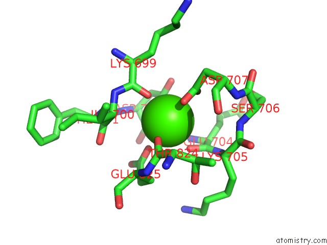
Mono view
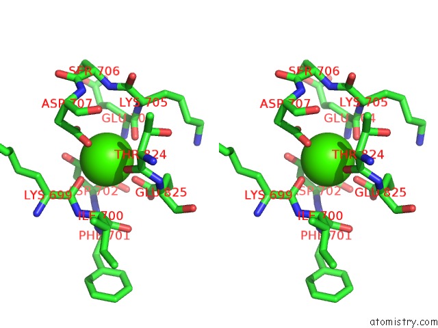
Stereo pair view
Mono view
Stereo pair view
A full contact list of Calcium with other atoms in the Ca binding
site number 1 of Crystal Structure of Streptococcus Pyogenes Endo-Beta-N- Acetylglucosaminidase (ENDOS2) within 5.0Å range:
|
Calcium binding site 2 out of 2 in 6e58
Go back to
Calcium binding site 2 out
of 2 in the Crystal Structure of Streptococcus Pyogenes Endo-Beta-N- Acetylglucosaminidase (ENDOS2)
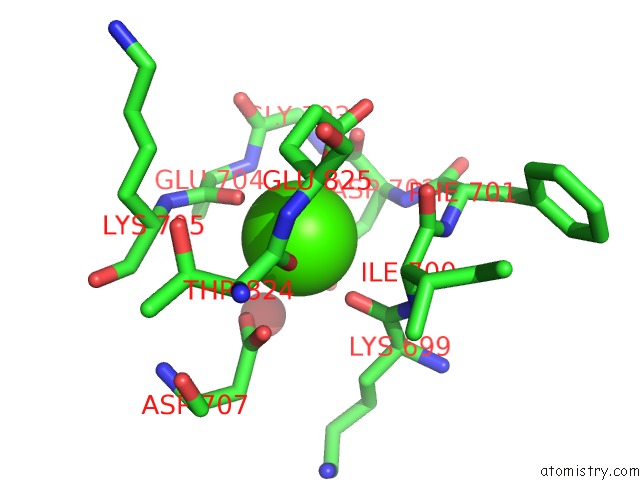
Mono view
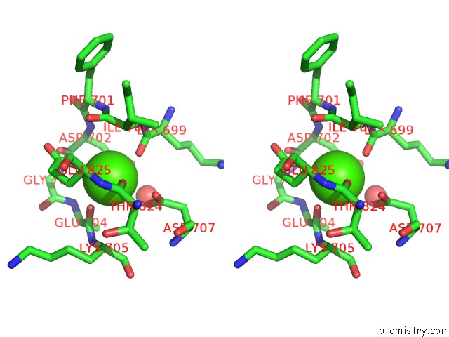
Stereo pair view
Mono view
Stereo pair view
A full contact list of Calcium with other atoms in the Ca binding
site number 2 of Crystal Structure of Streptococcus Pyogenes Endo-Beta-N- Acetylglucosaminidase (ENDOS2) within 5.0Å range:
|
Reference:
E.H.Klontz,
B.Trastoy,
D.Deredge,
J.K.Fields,
C.Li,
J.Orwenyo,
A.Marina,
R.Beadenkopf,
S.Gunther,
J.Flores,
P.L.Wintrode,
L.X.Wang,
M.E.Guerin,
E.J.Sundberg.
Molecular Basis of Broad Spectrumn-Glycan Specificity and Processing of Therapeutic Igg Monoclonal Antibodies By Endoglycosidase S2. Acs Cent Sci V. 5 524 2019.
ISSN: ESSN 2374-7943
PubMed: 30937380
DOI: 10.1021/ACSCENTSCI.8B00917
Page generated: Mon Jul 15 18:10:13 2024
ISSN: ESSN 2374-7943
PubMed: 30937380
DOI: 10.1021/ACSCENTSCI.8B00917
Last articles
Zn in 9J0NZn in 9J0O
Zn in 9J0P
Zn in 9FJX
Zn in 9EKB
Zn in 9C0F
Zn in 9CAH
Zn in 9CH0
Zn in 9CH3
Zn in 9CH1