Calcium »
PDB 6e54-6ela »
6ekn »
Calcium in PDB 6ekn: Crystal Structure of MMP12 in Complex with Inhibitor BE7.
Enzymatic activity of Crystal Structure of MMP12 in Complex with Inhibitor BE7.
All present enzymatic activity of Crystal Structure of MMP12 in Complex with Inhibitor BE7.:
3.4.24.65;
3.4.24.65;
Protein crystallography data
The structure of Crystal Structure of MMP12 in Complex with Inhibitor BE7., PDB code: 6ekn
was solved by
L.Ciccone,
L.Tepshi,
E.Nuti,
A.Rossello,
E.A.Stura,
with X-Ray Crystallography technique. A brief refinement statistics is given in the table below:
| Resolution Low / High (Å) | 36.74 / 1.20 |
| Space group | C 1 2 1 |
| Cell size a, b, c (Å), α, β, γ (°) | 51.560, 60.350, 54.560, 90.00, 116.10, 90.00 |
| R / Rfree (%) | 14.4 / 17.5 |
Other elements in 6ekn:
The structure of Crystal Structure of MMP12 in Complex with Inhibitor BE7. also contains other interesting chemical elements:
| Zinc | (Zn) | 2 atoms |
Calcium Binding Sites:
The binding sites of Calcium atom in the Crystal Structure of MMP12 in Complex with Inhibitor BE7.
(pdb code 6ekn). This binding sites where shown within
5.0 Angstroms radius around Calcium atom.
In total 3 binding sites of Calcium where determined in the Crystal Structure of MMP12 in Complex with Inhibitor BE7., PDB code: 6ekn:
Jump to Calcium binding site number: 1; 2; 3;
In total 3 binding sites of Calcium where determined in the Crystal Structure of MMP12 in Complex with Inhibitor BE7., PDB code: 6ekn:
Jump to Calcium binding site number: 1; 2; 3;
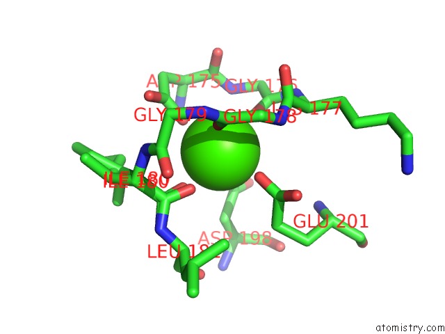
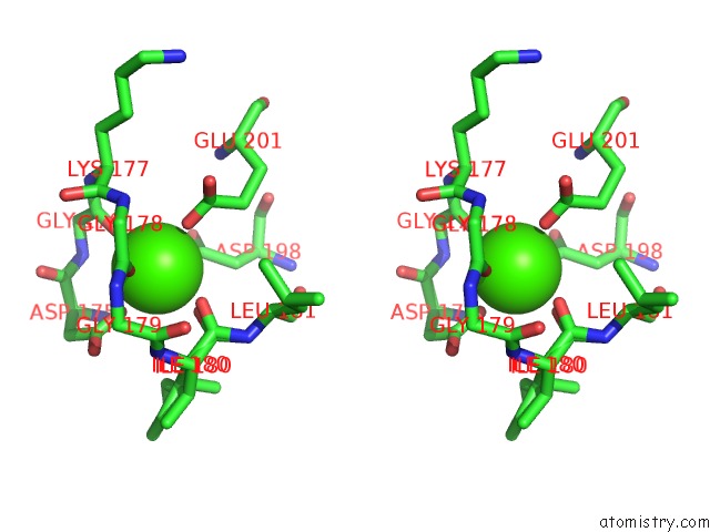
Calcium binding site 1 out of 3 in 6ekn
Go back to
Calcium binding site 1 out
of 3 in the Crystal Structure of MMP12 in Complex with Inhibitor BE7.
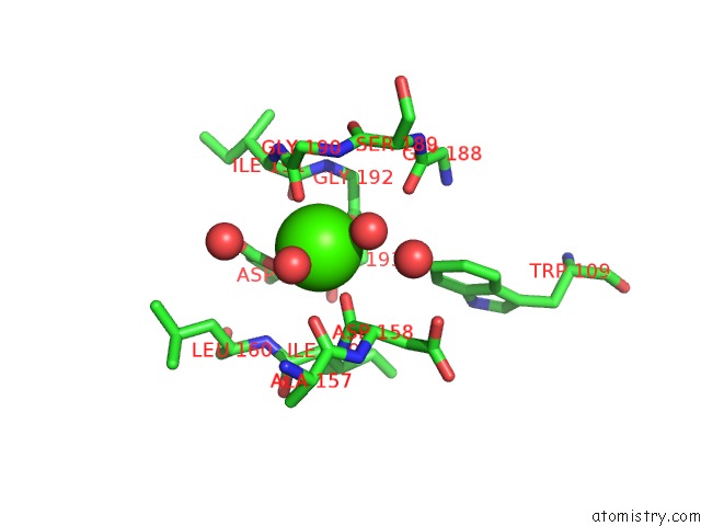
Mono view
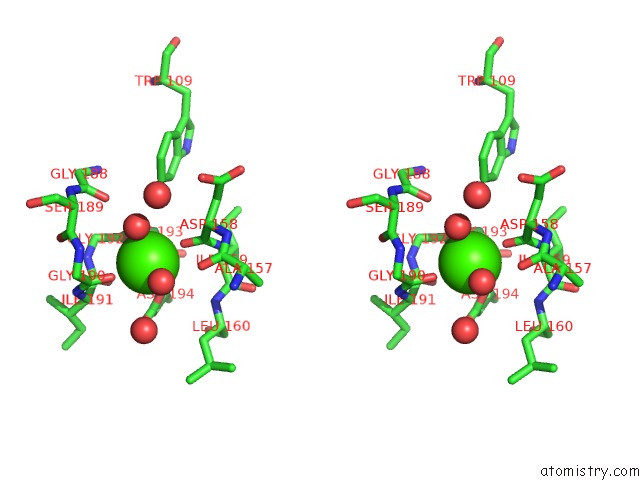
Stereo pair view
Mono view
Stereo pair view
A full contact list of Calcium with other atoms in the Ca binding
site number 1 of Crystal Structure of MMP12 in Complex with Inhibitor BE7. within 5.0Å range:
|
Calcium binding site 2 out of 3 in 6ekn
Go back to
Calcium binding site 2 out
of 3 in the Crystal Structure of MMP12 in Complex with Inhibitor BE7.
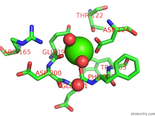
Mono view
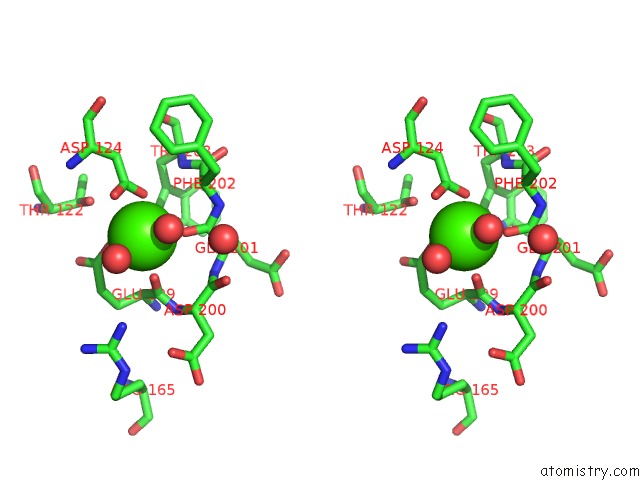
Stereo pair view
Mono view
Stereo pair view
A full contact list of Calcium with other atoms in the Ca binding
site number 2 of Crystal Structure of MMP12 in Complex with Inhibitor BE7. within 5.0Å range:
|
Calcium binding site 3 out of 3 in 6ekn
Go back to
Calcium binding site 3 out
of 3 in the Crystal Structure of MMP12 in Complex with Inhibitor BE7.
Mono view
Stereo pair view
Mono view
Stereo pair view
A full contact list of Calcium with other atoms in the Ca binding
site number 3 of Crystal Structure of MMP12 in Complex with Inhibitor BE7. within 5.0Å range:
|
Reference:
E.Nuti,
D.Cuffaro,
E.Bernardini,
C.Camodeca,
L.Panelli,
S.Chaves,
L.Ciccone,
L.Tepshi,
L.Vera,
E.Orlandini,
S.Nencetti,
E.A.Stura,
M.A.Santos,
V.Dive,
A.Rossello.
Development of Thioaryl-Based Matrix Metalloproteinase-12 Inhibitors with Alternative Zinc-Binding Groups: Synthesis, Potentiometric, uc(Nmr), and Crystallographic Studies. J. Med. Chem. V. 61 4421 2018.
ISSN: ISSN 1520-4804
PubMed: 29727184
DOI: 10.1021/ACS.JMEDCHEM.8B00096
Page generated: Mon Jul 15 18:21:45 2024
ISSN: ISSN 1520-4804
PubMed: 29727184
DOI: 10.1021/ACS.JMEDCHEM.8B00096
Last articles
Zn in 9J0NZn in 9J0O
Zn in 9J0P
Zn in 9FJX
Zn in 9EKB
Zn in 9C0F
Zn in 9CAH
Zn in 9CH0
Zn in 9CH3
Zn in 9CH1