Calcium »
PDB 6elt-6f2y »
6eqx »
Calcium in PDB 6eqx: X-Ray Structure of the Proprotein Convertase Furin Bound with the Competitive Inhibitor Arg-Arg-Arg-Val-Arg-Amba
Enzymatic activity of X-Ray Structure of the Proprotein Convertase Furin Bound with the Competitive Inhibitor Arg-Arg-Arg-Val-Arg-Amba
All present enzymatic activity of X-Ray Structure of the Proprotein Convertase Furin Bound with the Competitive Inhibitor Arg-Arg-Arg-Val-Arg-Amba:
3.4.21.75;
3.4.21.75;
Protein crystallography data
The structure of X-Ray Structure of the Proprotein Convertase Furin Bound with the Competitive Inhibitor Arg-Arg-Arg-Val-Arg-Amba, PDB code: 6eqx
was solved by
S.O.Dahms,
M.E.Than,
with X-Ray Crystallography technique. A brief refinement statistics is given in the table below:
| Resolution Low / High (Å) | 47.16 / 1.99 |
| Space group | P 65 2 2 |
| Cell size a, b, c (Å), α, β, γ (°) | 131.805, 131.805, 155.347, 90.00, 90.00, 120.00 |
| R / Rfree (%) | 16.2 / 18.4 |
Other elements in 6eqx:
The structure of X-Ray Structure of the Proprotein Convertase Furin Bound with the Competitive Inhibitor Arg-Arg-Arg-Val-Arg-Amba also contains other interesting chemical elements:
| Chlorine | (Cl) | 1 atom |
| Sodium | (Na) | 4 atoms |
Calcium Binding Sites:
The binding sites of Calcium atom in the X-Ray Structure of the Proprotein Convertase Furin Bound with the Competitive Inhibitor Arg-Arg-Arg-Val-Arg-Amba
(pdb code 6eqx). This binding sites where shown within
5.0 Angstroms radius around Calcium atom.
In total 3 binding sites of Calcium where determined in the X-Ray Structure of the Proprotein Convertase Furin Bound with the Competitive Inhibitor Arg-Arg-Arg-Val-Arg-Amba, PDB code: 6eqx:
Jump to Calcium binding site number: 1; 2; 3;
In total 3 binding sites of Calcium where determined in the X-Ray Structure of the Proprotein Convertase Furin Bound with the Competitive Inhibitor Arg-Arg-Arg-Val-Arg-Amba, PDB code: 6eqx:
Jump to Calcium binding site number: 1; 2; 3;
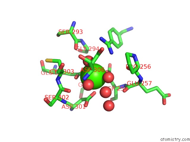
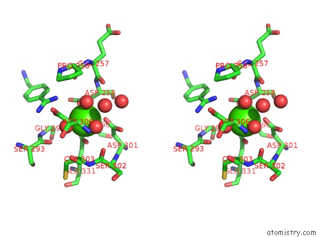
Calcium binding site 1 out of 3 in 6eqx
Go back to
Calcium binding site 1 out
of 3 in the X-Ray Structure of the Proprotein Convertase Furin Bound with the Competitive Inhibitor Arg-Arg-Arg-Val-Arg-Amba
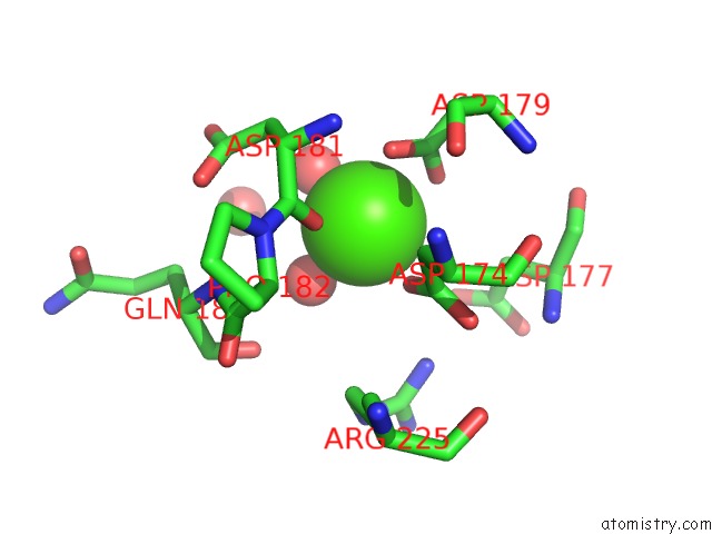
Mono view
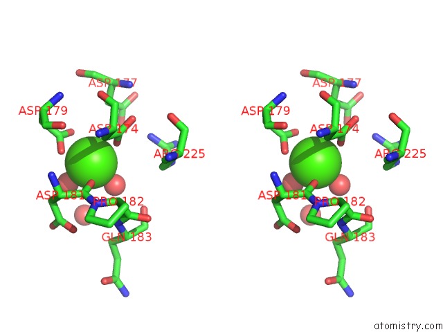
Stereo pair view
Mono view
Stereo pair view
A full contact list of Calcium with other atoms in the Ca binding
site number 1 of X-Ray Structure of the Proprotein Convertase Furin Bound with the Competitive Inhibitor Arg-Arg-Arg-Val-Arg-Amba within 5.0Å range:
|
Calcium binding site 2 out of 3 in 6eqx
Go back to
Calcium binding site 2 out
of 3 in the X-Ray Structure of the Proprotein Convertase Furin Bound with the Competitive Inhibitor Arg-Arg-Arg-Val-Arg-Amba
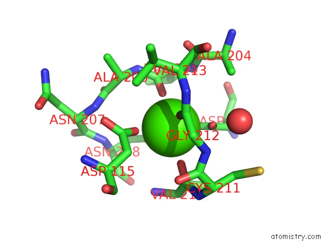
Mono view
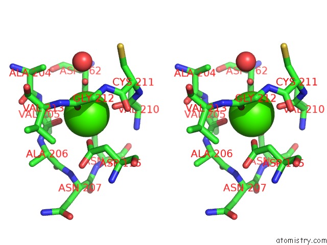
Stereo pair view
Mono view
Stereo pair view
A full contact list of Calcium with other atoms in the Ca binding
site number 2 of X-Ray Structure of the Proprotein Convertase Furin Bound with the Competitive Inhibitor Arg-Arg-Arg-Val-Arg-Amba within 5.0Å range:
|
Calcium binding site 3 out of 3 in 6eqx
Go back to
Calcium binding site 3 out
of 3 in the X-Ray Structure of the Proprotein Convertase Furin Bound with the Competitive Inhibitor Arg-Arg-Arg-Val-Arg-Amba
Mono view
Stereo pair view
Mono view
Stereo pair view
A full contact list of Calcium with other atoms in the Ca binding
site number 3 of X-Ray Structure of the Proprotein Convertase Furin Bound with the Competitive Inhibitor Arg-Arg-Arg-Val-Arg-Amba within 5.0Å range:
|
Reference:
S.O.Dahms,
K.Hardes,
T.Steinmetzer,
M.E.Than.
X-Ray Structures of the Proprotein Convertase Furin Bound with Substrate Analogue Inhibitors Reveal Substrate Specificity Determinants Beyond the S4 Pocket. Biochemistry V. 57 925 2018.
ISSN: ISSN 1520-4995
PubMed: 29314830
DOI: 10.1021/ACS.BIOCHEM.7B01124
Page generated: Mon Jul 15 18:37:01 2024
ISSN: ISSN 1520-4995
PubMed: 29314830
DOI: 10.1021/ACS.BIOCHEM.7B01124
Last articles
Zn in 9J0NZn in 9J0O
Zn in 9J0P
Zn in 9FJX
Zn in 9EKB
Zn in 9C0F
Zn in 9CAH
Zn in 9CH0
Zn in 9CH3
Zn in 9CH1