Calcium »
PDB 6elt-6f2y »
6f1j »
Calcium in PDB 6f1j: Structure of A Talaromyces Pinophilus GH62 Arabinofuranosidase in Complex with Aradnj at 1.25A Resolution
Protein crystallography data
The structure of Structure of A Talaromyces Pinophilus GH62 Arabinofuranosidase in Complex with Aradnj at 1.25A Resolution, PDB code: 6f1j
was solved by
O.V.Moroz,
L.Sobala,
E.Blagova,
T.Coyle,
K.B.R.Morkeberg Krogh,
P.Wei,
K.Stubbs,
K.S.Wilson,
G.J.Davies,
with X-Ray Crystallography technique. A brief refinement statistics is given in the table below:
| Resolution Low / High (Å) | 33.52 / 1.25 |
| Space group | P 1 21 1 |
| Cell size a, b, c (Å), α, β, γ (°) | 43.827, 88.978, 72.663, 90.00, 95.22, 90.00 |
| R / Rfree (%) | 12 / 13.6 |
Other elements in 6f1j:
The structure of Structure of A Talaromyces Pinophilus GH62 Arabinofuranosidase in Complex with Aradnj at 1.25A Resolution also contains other interesting chemical elements:
| Zinc | (Zn) | 2 atoms |
Calcium Binding Sites:
The binding sites of Calcium atom in the Structure of A Talaromyces Pinophilus GH62 Arabinofuranosidase in Complex with Aradnj at 1.25A Resolution
(pdb code 6f1j). This binding sites where shown within
5.0 Angstroms radius around Calcium atom.
In total 2 binding sites of Calcium where determined in the Structure of A Talaromyces Pinophilus GH62 Arabinofuranosidase in Complex with Aradnj at 1.25A Resolution, PDB code: 6f1j:
Jump to Calcium binding site number: 1; 2;
In total 2 binding sites of Calcium where determined in the Structure of A Talaromyces Pinophilus GH62 Arabinofuranosidase in Complex with Aradnj at 1.25A Resolution, PDB code: 6f1j:
Jump to Calcium binding site number: 1; 2;
Calcium binding site 1 out of 2 in 6f1j
Go back to
Calcium binding site 1 out
of 2 in the Structure of A Talaromyces Pinophilus GH62 Arabinofuranosidase in Complex with Aradnj at 1.25A Resolution
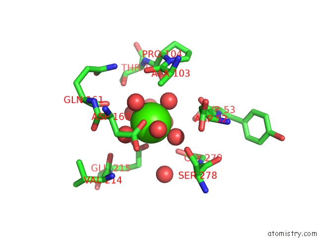
Mono view
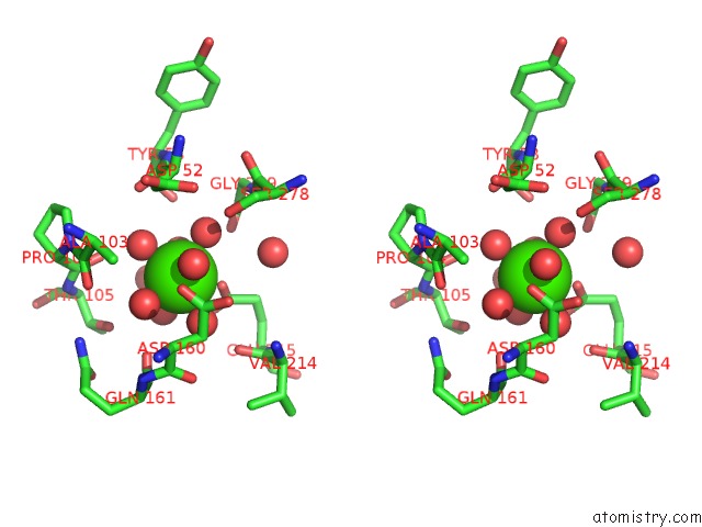
Stereo pair view
Mono view
Stereo pair view
A full contact list of Calcium with other atoms in the Ca binding
site number 1 of Structure of A Talaromyces Pinophilus GH62 Arabinofuranosidase in Complex with Aradnj at 1.25A Resolution within 5.0Å range:
|
Calcium binding site 2 out of 2 in 6f1j
Go back to
Calcium binding site 2 out
of 2 in the Structure of A Talaromyces Pinophilus GH62 Arabinofuranosidase in Complex with Aradnj at 1.25A Resolution
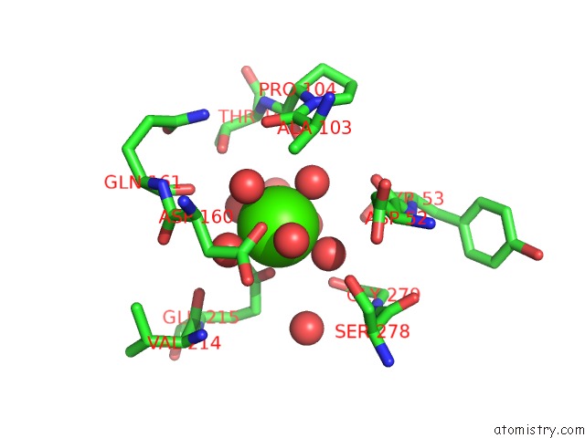
Mono view
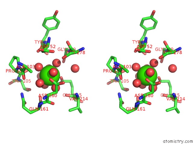
Stereo pair view
Mono view
Stereo pair view
A full contact list of Calcium with other atoms in the Ca binding
site number 2 of Structure of A Talaromyces Pinophilus GH62 Arabinofuranosidase in Complex with Aradnj at 1.25A Resolution within 5.0Å range:
|
Reference:
O.V.Moroz,
L.F.Sobala,
E.Blagova,
T.Coyle,
W.Peng,
K.B.R.Morkeberg Krogh,
K.A.Stubbs,
K.S.Wilson,
G.J.Davies.
Structure of A Talaromyces Pinophilus GH62 Arabinofuranosidase in Complex with Aradnj at 1.25 Angstrom Resolution. Acta Crystallogr F Struct V. 74 490 2018BIOL Commun.
ISSN: ESSN 2053-230X
PubMed: 30084398
DOI: 10.1107/S2053230X18000250
Page generated: Wed Jul 9 13:57:02 2025
ISSN: ESSN 2053-230X
PubMed: 30084398
DOI: 10.1107/S2053230X18000250
Last articles
F in 4DL8F in 4DK7
F in 4DKR
F in 4DKP
F in 4DKQ
F in 4DK8
F in 4DKO
F in 4DKJ
F in 4DHP
F in 4DBU