Calcium »
PDB 6f30-6fi4 »
6f4k »
Calcium in PDB 6f4k: Crystal Structure of Glutathione Transferase Omega 3S From Trametes Versicolor in Complex with Hexyl-Glutathione
Protein crystallography data
The structure of Crystal Structure of Glutathione Transferase Omega 3S From Trametes Versicolor in Complex with Hexyl-Glutathione, PDB code: 6f4k
was solved by
M.Schwartz,
F.Favier,
C.Didierjean,
with X-Ray Crystallography technique. A brief refinement statistics is given in the table below:
| Resolution Low / High (Å) | 47.80 / 1.55 |
| Space group | P 21 21 21 |
| Cell size a, b, c (Å), α, β, γ (°) | 50.530, 104.140, 107.590, 90.00, 90.00, 90.00 |
| R / Rfree (%) | 16.1 / 18.4 |
Calcium Binding Sites:
The binding sites of Calcium atom in the Crystal Structure of Glutathione Transferase Omega 3S From Trametes Versicolor in Complex with Hexyl-Glutathione
(pdb code 6f4k). This binding sites where shown within
5.0 Angstroms radius around Calcium atom.
In total only one binding site of Calcium was determined in the Crystal Structure of Glutathione Transferase Omega 3S From Trametes Versicolor in Complex with Hexyl-Glutathione, PDB code: 6f4k:
In total only one binding site of Calcium was determined in the Crystal Structure of Glutathione Transferase Omega 3S From Trametes Versicolor in Complex with Hexyl-Glutathione, PDB code: 6f4k:
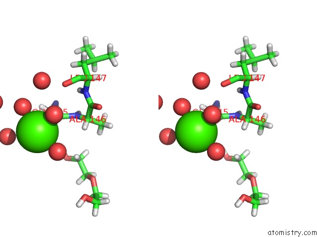
Calcium binding site 1 out of 1 in 6f4k
Go back to
Calcium binding site 1 out
of 1 in the Crystal Structure of Glutathione Transferase Omega 3S From Trametes Versicolor in Complex with Hexyl-Glutathione
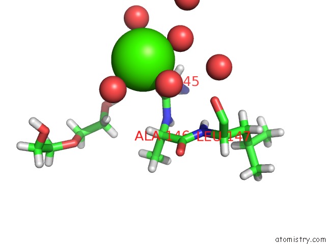
Mono view
Stereo pair view
Mono view
Stereo pair view
A full contact list of Calcium with other atoms in the Ca binding
site number 1 of Crystal Structure of Glutathione Transferase Omega 3S From Trametes Versicolor in Complex with Hexyl-Glutathione within 5.0Å range:
|
Reference:
M.Schwartz,
T.Perrot,
E.Aubert,
S.Dumarcay,
F.Favier,
P.Gerardin,
M.Morel-Rouhier,
G.Mulliert,
F.Saiag,
C.Didierjean,
E.Gelhaye.
Molecular Recognition of Wood Polyphenols By Phase II Detoxification Enzymes of the White Rot Trametes Versicolor. Sci Rep V. 8 8472 2018.
ISSN: ESSN 2045-2322
PubMed: 29855494
DOI: 10.1038/S41598-018-26601-3
Page generated: Wed Jul 9 14:06:01 2025
ISSN: ESSN 2045-2322
PubMed: 29855494
DOI: 10.1038/S41598-018-26601-3
Last articles
Ca in 7M66Ca in 7M8C
Ca in 7M8B
Ca in 7M8T
Ca in 7M8A
Ca in 7M89
Ca in 7M88
Ca in 7M87
Ca in 7M86
Ca in 7M85