Calcium »
PDB 6f30-6fi4 »
6fat »
Calcium in PDB 6fat: The Crystal Structure of A Feruloyl Esterase C From Fusarium Oxysporum.
Protein crystallography data
The structure of The Crystal Structure of A Feruloyl Esterase C From Fusarium Oxysporum., PDB code: 6fat
was solved by
M.Dimarogona,
E.D.Chrysina,
with X-Ray Crystallography technique. A brief refinement statistics is given in the table below:
| Resolution Low / High (Å) | 102.35 / 2.30 |
| Space group | P 1 21 1 |
| Cell size a, b, c (Å), α, β, γ (°) | 67.536, 87.467, 106.565, 90.00, 106.18, 90.00 |
| R / Rfree (%) | 20.7 / 23.5 |
Calcium Binding Sites:
The binding sites of Calcium atom in the The Crystal Structure of A Feruloyl Esterase C From Fusarium Oxysporum.
(pdb code 6fat). This binding sites where shown within
5.0 Angstroms radius around Calcium atom.
In total 2 binding sites of Calcium where determined in the The Crystal Structure of A Feruloyl Esterase C From Fusarium Oxysporum., PDB code: 6fat:
Jump to Calcium binding site number: 1; 2;
In total 2 binding sites of Calcium where determined in the The Crystal Structure of A Feruloyl Esterase C From Fusarium Oxysporum., PDB code: 6fat:
Jump to Calcium binding site number: 1; 2;
Calcium binding site 1 out of 2 in 6fat
Go back to
Calcium binding site 1 out
of 2 in the The Crystal Structure of A Feruloyl Esterase C From Fusarium Oxysporum.
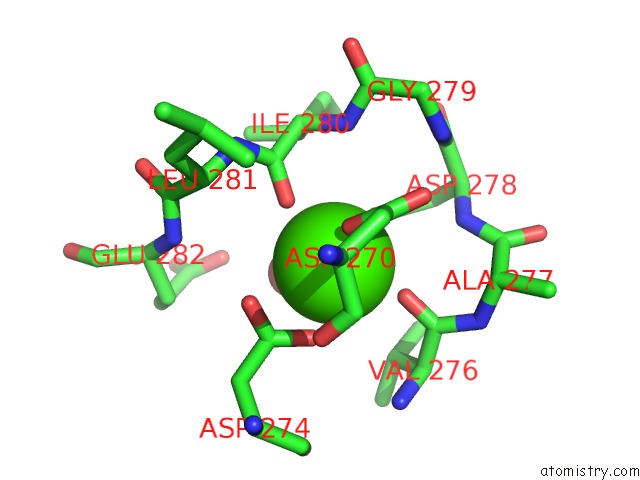
Mono view
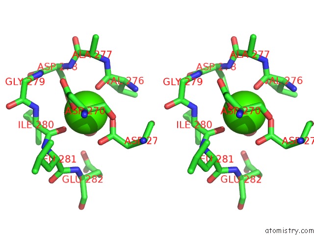
Stereo pair view
Mono view
Stereo pair view
A full contact list of Calcium with other atoms in the Ca binding
site number 1 of The Crystal Structure of A Feruloyl Esterase C From Fusarium Oxysporum. within 5.0Å range:
|
Calcium binding site 2 out of 2 in 6fat
Go back to
Calcium binding site 2 out
of 2 in the The Crystal Structure of A Feruloyl Esterase C From Fusarium Oxysporum.
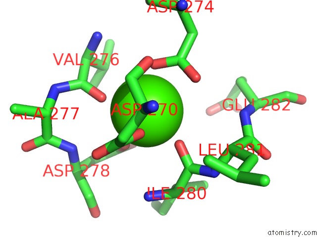
Mono view
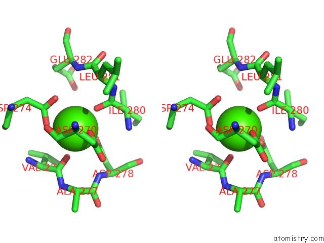
Stereo pair view
Mono view
Stereo pair view
A full contact list of Calcium with other atoms in the Ca binding
site number 2 of The Crystal Structure of A Feruloyl Esterase C From Fusarium Oxysporum. within 5.0Å range:
|
Reference:
M.Dimarogona,
E.D.Chrysina.
The Crystal Structure of A Feruloyl Esterase C From Fusarium Oxysporum. To Be Published.
Page generated: Wed Jul 9 14:08:54 2025
Last articles
Ca in 7MMPCa in 7MMS
Ca in 7MJR
Ca in 7MIY
Ca in 7MIX
Ca in 7MGD
Ca in 7MGC
Ca in 7MIS
Ca in 7MGB
Ca in 7MIR