Calcium »
PDB 6f30-6fi4 »
6fb0 »
Calcium in PDB 6fb0: Crystal Structure of A Tailored I-Crei Homing Endonuclease Protein (3115 Variant) in Complex with Its Target Dna (Haemoglobin Beta Subunit Gene) in the Presence of Calcium
Protein crystallography data
The structure of Crystal Structure of A Tailored I-Crei Homing Endonuclease Protein (3115 Variant) in Complex with Its Target Dna (Haemoglobin Beta Subunit Gene) in the Presence of Calcium, PDB code: 6fb0
was solved by
R.Molina,
J.Prieto,
with X-Ray Crystallography technique. A brief refinement statistics is given in the table below:
| Resolution Low / High (Å) | 33.84 / 2.15 |
| Space group | P 1 21 1 |
| Cell size a, b, c (Å), α, β, γ (°) | 45.437, 67.673, 83.525, 90.00, 96.75, 90.00 |
| R / Rfree (%) | 17.6 / 22.9 |
Calcium Binding Sites:
The binding sites of Calcium atom in the Crystal Structure of A Tailored I-Crei Homing Endonuclease Protein (3115 Variant) in Complex with Its Target Dna (Haemoglobin Beta Subunit Gene) in the Presence of Calcium
(pdb code 6fb0). This binding sites where shown within
5.0 Angstroms radius around Calcium atom.
In total 4 binding sites of Calcium where determined in the Crystal Structure of A Tailored I-Crei Homing Endonuclease Protein (3115 Variant) in Complex with Its Target Dna (Haemoglobin Beta Subunit Gene) in the Presence of Calcium, PDB code: 6fb0:
Jump to Calcium binding site number: 1; 2; 3; 4;
In total 4 binding sites of Calcium where determined in the Crystal Structure of A Tailored I-Crei Homing Endonuclease Protein (3115 Variant) in Complex with Its Target Dna (Haemoglobin Beta Subunit Gene) in the Presence of Calcium, PDB code: 6fb0:
Jump to Calcium binding site number: 1; 2; 3; 4;
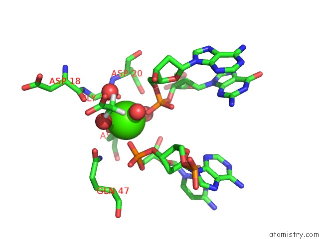
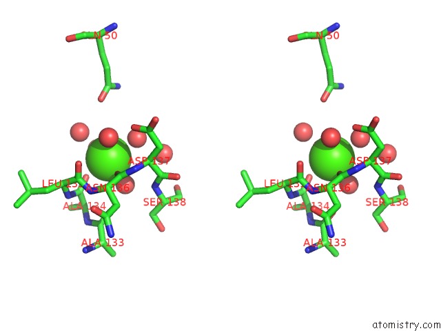
Calcium binding site 1 out of 4 in 6fb0
Go back to
Calcium binding site 1 out
of 4 in the Crystal Structure of A Tailored I-Crei Homing Endonuclease Protein (3115 Variant) in Complex with Its Target Dna (Haemoglobin Beta Subunit Gene) in the Presence of Calcium
Mono view
Stereo pair view
Mono view
Stereo pair view
A full contact list of Calcium with other atoms in the Ca binding
site number 1 of Crystal Structure of A Tailored I-Crei Homing Endonuclease Protein (3115 Variant) in Complex with Its Target Dna (Haemoglobin Beta Subunit Gene) in the Presence of Calcium within 5.0Å range:
|
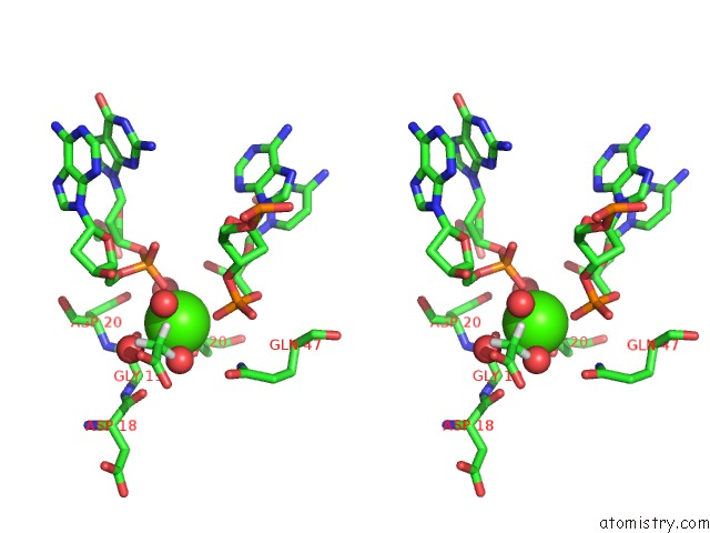
Calcium binding site 2 out of 4 in 6fb0
Go back to
Calcium binding site 2 out
of 4 in the Crystal Structure of A Tailored I-Crei Homing Endonuclease Protein (3115 Variant) in Complex with Its Target Dna (Haemoglobin Beta Subunit Gene) in the Presence of Calcium

Mono view
Stereo pair view
Mono view
Stereo pair view
A full contact list of Calcium with other atoms in the Ca binding
site number 2 of Crystal Structure of A Tailored I-Crei Homing Endonuclease Protein (3115 Variant) in Complex with Its Target Dna (Haemoglobin Beta Subunit Gene) in the Presence of Calcium within 5.0Å range:
|
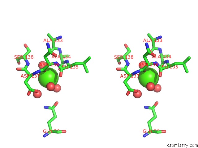
Calcium binding site 3 out of 4 in 6fb0
Go back to
Calcium binding site 3 out
of 4 in the Crystal Structure of A Tailored I-Crei Homing Endonuclease Protein (3115 Variant) in Complex with Its Target Dna (Haemoglobin Beta Subunit Gene) in the Presence of Calcium

Mono view
Stereo pair view
Mono view
Stereo pair view
A full contact list of Calcium with other atoms in the Ca binding
site number 3 of Crystal Structure of A Tailored I-Crei Homing Endonuclease Protein (3115 Variant) in Complex with Its Target Dna (Haemoglobin Beta Subunit Gene) in the Presence of Calcium within 5.0Å range:
|
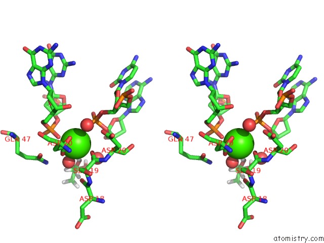
Calcium binding site 4 out of 4 in 6fb0
Go back to
Calcium binding site 4 out
of 4 in the Crystal Structure of A Tailored I-Crei Homing Endonuclease Protein (3115 Variant) in Complex with Its Target Dna (Haemoglobin Beta Subunit Gene) in the Presence of Calcium

Mono view
Stereo pair view
Mono view
Stereo pair view
A full contact list of Calcium with other atoms in the Ca binding
site number 4 of Crystal Structure of A Tailored I-Crei Homing Endonuclease Protein (3115 Variant) in Complex with Its Target Dna (Haemoglobin Beta Subunit Gene) in the Presence of Calcium within 5.0Å range:
|
Reference:
J.Prieto,
P.Redondo,
B.Lopez-Mendez,
M.D'abramo,
N.Merino,
F.J.Blanco,
P.Duchateau,
G.Montoya,
R.Molina.
Understanding the Indirect Dna Read-Out Specificity of I-Crei Meganuclease. Sci Rep V. 8 10286 2018.
ISSN: ESSN 2045-2322
PubMed: 29980759
DOI: 10.1038/S41598-018-28599-0
Page generated: Wed Jul 9 14:09:14 2025
ISSN: ESSN 2045-2322
PubMed: 29980759
DOI: 10.1038/S41598-018-28599-0
Last articles
Cl in 5G54Cl in 5G4A
Cl in 5G4Q
Cl in 5G47
Cl in 5G42
Cl in 5G3S
Cl in 5G2P
Cl in 5G2T
Cl in 5G36
Cl in 5G2D