Calcium »
PDB 6f30-6fi4 »
6fc2 »
Calcium in PDB 6fc2: Crystal Structure of the EIF4E-EAP1P Complex From Saccharomyces Cerevisiae
Protein crystallography data
The structure of Crystal Structure of the EIF4E-EAP1P Complex From Saccharomyces Cerevisiae, PDB code: 6fc2
was solved by
S.Gruener,
E.Valkov,
with X-Ray Crystallography technique. A brief refinement statistics is given in the table below:
| Resolution Low / High (Å) | 48.26 / 1.92 |
| Space group | P 21 21 2 |
| Cell size a, b, c (Å), α, β, γ (°) | 87.574, 95.116, 56.009, 90.00, 90.00, 90.00 |
| R / Rfree (%) | 17.4 / 20.7 |
Calcium Binding Sites:
The binding sites of Calcium atom in the Crystal Structure of the EIF4E-EAP1P Complex From Saccharomyces Cerevisiae
(pdb code 6fc2). This binding sites where shown within
5.0 Angstroms radius around Calcium atom.
In total only one binding site of Calcium was determined in the Crystal Structure of the EIF4E-EAP1P Complex From Saccharomyces Cerevisiae, PDB code: 6fc2:
In total only one binding site of Calcium was determined in the Crystal Structure of the EIF4E-EAP1P Complex From Saccharomyces Cerevisiae, PDB code: 6fc2:
Calcium binding site 1 out of 1 in 6fc2
Go back to
Calcium binding site 1 out
of 1 in the Crystal Structure of the EIF4E-EAP1P Complex From Saccharomyces Cerevisiae
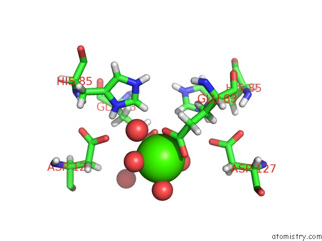
Mono view
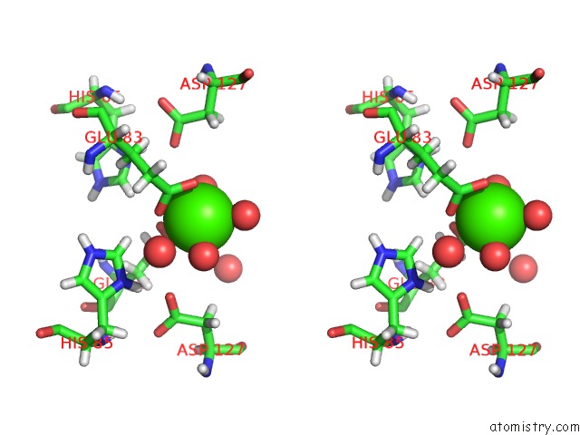
Stereo pair view
Mono view
Stereo pair view
A full contact list of Calcium with other atoms in the Ca binding
site number 1 of Crystal Structure of the EIF4E-EAP1P Complex From Saccharomyces Cerevisiae within 5.0Å range:
|
Reference:
S.Gruner,
R.Weber,
D.Peter,
M.Y.Chung,
C.Igreja,
E.Valkov,
E.Izaurralde.
Structural Motifs in EIF4G and 4E-Bps Modulate Their Binding to EIF4E to Regulate Translation Initiation in Yeast. Nucleic Acids Res. V. 46 6893 2018.
ISSN: ESSN 1362-4962
PubMed: 30053226
DOI: 10.1093/NAR/GKY542
Page generated: Wed Jul 9 14:09:36 2025
ISSN: ESSN 1362-4962
PubMed: 30053226
DOI: 10.1093/NAR/GKY542
Last articles
Ca in 7ONGCa in 7OR0
Ca in 7OP2
Ca in 7OO7
Ca in 7OO5
Ca in 7O85
Ca in 7OIH
Ca in 7ONA
Ca in 7OMG
Ca in 7OKW