Calcium »
PDB 6f30-6fi4 »
6fgc »
Calcium in PDB 6fgc: Crystal Structure of Gephyrin E Domain in Complex with Artesunate
Enzymatic activity of Crystal Structure of Gephyrin E Domain in Complex with Artesunate
All present enzymatic activity of Crystal Structure of Gephyrin E Domain in Complex with Artesunate:
2.10.1.1; 2.7.7.75;
2.10.1.1; 2.7.7.75;
Protein crystallography data
The structure of Crystal Structure of Gephyrin E Domain in Complex with Artesunate, PDB code: 6fgc
was solved by
V.B.Kasaragod,
H.Schindelin,
with X-Ray Crystallography technique. A brief refinement statistics is given in the table below:
| Resolution Low / High (Å) | 43.55 / 1.50 |
| Space group | I 2 2 2 |
| Cell size a, b, c (Å), α, β, γ (°) | 87.100, 99.220, 113.260, 90.00, 90.00, 90.00 |
| R / Rfree (%) | 14.3 / 16.2 |
Other elements in 6fgc:
The structure of Crystal Structure of Gephyrin E Domain in Complex with Artesunate also contains other interesting chemical elements:
| Chlorine | (Cl) | 1 atom |
Calcium Binding Sites:
The binding sites of Calcium atom in the Crystal Structure of Gephyrin E Domain in Complex with Artesunate
(pdb code 6fgc). This binding sites where shown within
5.0 Angstroms radius around Calcium atom.
In total only one binding site of Calcium was determined in the Crystal Structure of Gephyrin E Domain in Complex with Artesunate, PDB code: 6fgc:
In total only one binding site of Calcium was determined in the Crystal Structure of Gephyrin E Domain in Complex with Artesunate, PDB code: 6fgc:
Calcium binding site 1 out of 1 in 6fgc
Go back to
Calcium binding site 1 out
of 1 in the Crystal Structure of Gephyrin E Domain in Complex with Artesunate
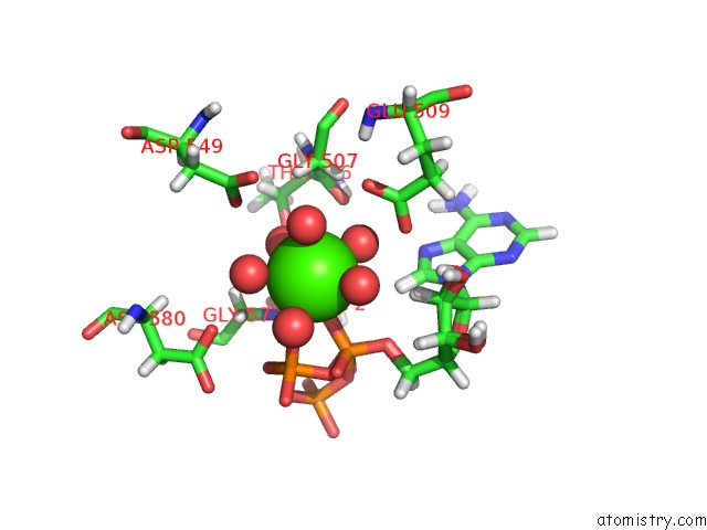
Mono view
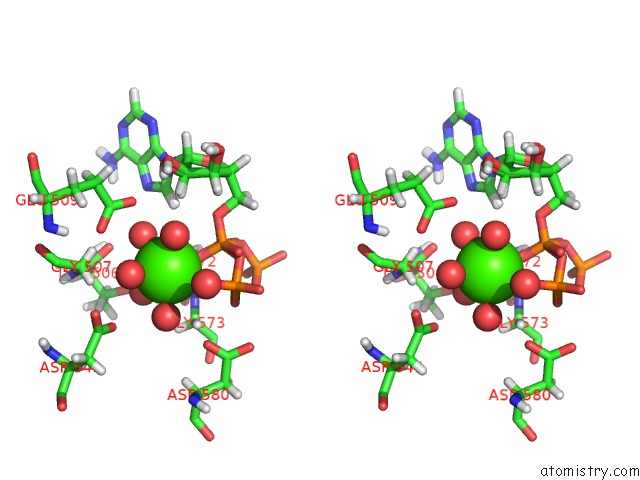
Stereo pair view
Mono view
Stereo pair view
A full contact list of Calcium with other atoms in the Ca binding
site number 1 of Crystal Structure of Gephyrin E Domain in Complex with Artesunate within 5.0Å range:
|
Reference:
V.B.Kasaragod,
T.J.Hausrat,
N.Schaefer,
M.Kuhn,
N.R.Christensen,
I.Tessmer,
H.M.Maric,
K.L.Madsen,
C.Sotriffer,
C.Villmann,
M.Kneussel,
H.Schindelin.
Elucidating the Molecular Basis For Inhibitory Neurotransmission Regulation By Artemisinins. Neuron V. 101 673 2019.
ISSN: ISSN 1097-4199
PubMed: 30704910
DOI: 10.1016/J.NEURON.2019.01.001
Page generated: Wed Jul 9 14:10:23 2025
ISSN: ISSN 1097-4199
PubMed: 30704910
DOI: 10.1016/J.NEURON.2019.01.001
Last articles
Fe in 2YXOFe in 2YRS
Fe in 2YXC
Fe in 2YNM
Fe in 2YVJ
Fe in 2YP1
Fe in 2YU2
Fe in 2YU1
Fe in 2YQB
Fe in 2YOO