Calcium »
PDB 6fi8-6fzv »
6fwk »
Calcium in PDB 6fwk: The Crystal Structure of POL2CORE-M644G in Complex with Dna and An Incoming Nucleotide
Enzymatic activity of The Crystal Structure of POL2CORE-M644G in Complex with Dna and An Incoming Nucleotide
All present enzymatic activity of The Crystal Structure of POL2CORE-M644G in Complex with Dna and An Incoming Nucleotide:
2.7.7.7;
2.7.7.7;
Protein crystallography data
The structure of The Crystal Structure of POL2CORE-M644G in Complex with Dna and An Incoming Nucleotide, PDB code: 6fwk
was solved by
V.Parkash,
E.Johansson,
with X-Ray Crystallography technique. A brief refinement statistics is given in the table below:
| Resolution Low / High (Å) | 19.99 / 2.50 |
| Space group | P 1 2 1 |
| Cell size a, b, c (Å), α, β, γ (°) | 153.961, 70.342, 158.965, 90.00, 112.82, 90.00 |
| R / Rfree (%) | 22.3 / 26.3 |
Other elements in 6fwk:
The structure of The Crystal Structure of POL2CORE-M644G in Complex with Dna and An Incoming Nucleotide also contains other interesting chemical elements:
| Iron | (Fe) | 2 atoms |
Calcium Binding Sites:
The binding sites of Calcium atom in the The Crystal Structure of POL2CORE-M644G in Complex with Dna and An Incoming Nucleotide
(pdb code 6fwk). This binding sites where shown within
5.0 Angstroms radius around Calcium atom.
In total 5 binding sites of Calcium where determined in the The Crystal Structure of POL2CORE-M644G in Complex with Dna and An Incoming Nucleotide, PDB code: 6fwk:
Jump to Calcium binding site number: 1; 2; 3; 4; 5;
In total 5 binding sites of Calcium where determined in the The Crystal Structure of POL2CORE-M644G in Complex with Dna and An Incoming Nucleotide, PDB code: 6fwk:
Jump to Calcium binding site number: 1; 2; 3; 4; 5;
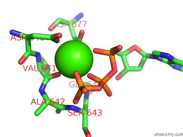
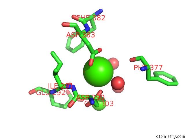
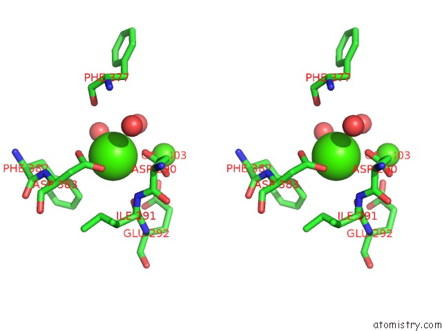
Calcium binding site 1 out of 5 in 6fwk
Go back to
Calcium binding site 1 out
of 5 in the The Crystal Structure of POL2CORE-M644G in Complex with Dna and An Incoming Nucleotide
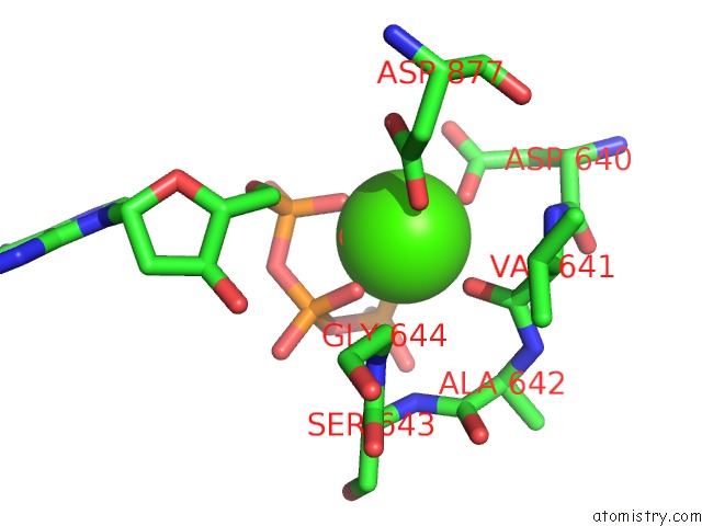
Mono view
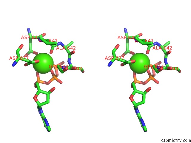
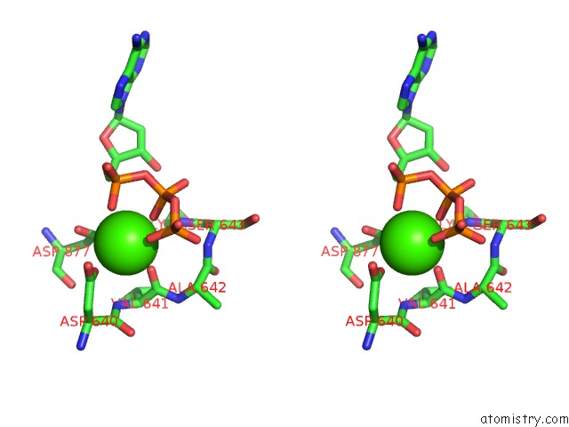
Stereo pair view
Mono view
Stereo pair view
A full contact list of Calcium with other atoms in the Ca binding
site number 1 of The Crystal Structure of POL2CORE-M644G in Complex with Dna and An Incoming Nucleotide within 5.0Å range:
|
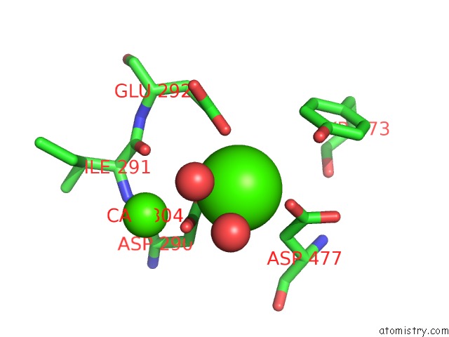
Calcium binding site 2 out of 5 in 6fwk
Go back to
Calcium binding site 2 out
of 5 in the The Crystal Structure of POL2CORE-M644G in Complex with Dna and An Incoming Nucleotide
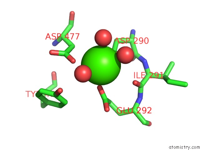
Mono view
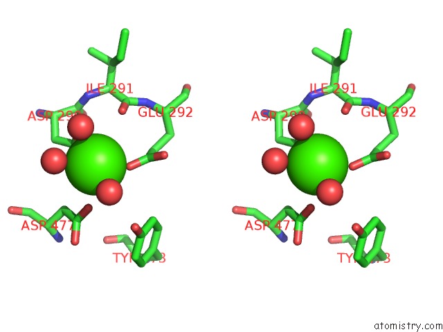
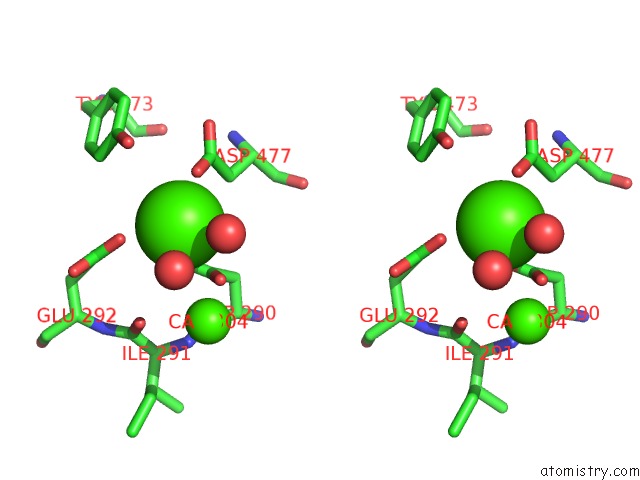
Stereo pair view
Mono view
Stereo pair view
A full contact list of Calcium with other atoms in the Ca binding
site number 2 of The Crystal Structure of POL2CORE-M644G in Complex with Dna and An Incoming Nucleotide within 5.0Å range:
|
Calcium binding site 3 out of 5 in 6fwk
Go back to
Calcium binding site 3 out
of 5 in the The Crystal Structure of POL2CORE-M644G in Complex with Dna and An Incoming Nucleotide
Mono view
Stereo pair view
Mono view
Stereo pair view
A full contact list of Calcium with other atoms in the Ca binding
site number 3 of The Crystal Structure of POL2CORE-M644G in Complex with Dna and An Incoming Nucleotide within 5.0Å range:
|
Calcium binding site 4 out of 5 in 6fwk
Go back to
Calcium binding site 4 out
of 5 in the The Crystal Structure of POL2CORE-M644G in Complex with Dna and An Incoming Nucleotide
Mono view
Stereo pair view
Mono view
Stereo pair view
A full contact list of Calcium with other atoms in the Ca binding
site number 4 of The Crystal Structure of POL2CORE-M644G in Complex with Dna and An Incoming Nucleotide within 5.0Å range:
|
Calcium binding site 5 out of 5 in 6fwk
Go back to
Calcium binding site 5 out
of 5 in the The Crystal Structure of POL2CORE-M644G in Complex with Dna and An Incoming Nucleotide
Mono view
Stereo pair view
Mono view
Stereo pair view
A full contact list of Calcium with other atoms in the Ca binding
site number 5 of The Crystal Structure of POL2CORE-M644G in Complex with Dna and An Incoming Nucleotide within 5.0Å range:
|
Reference:
V.Parkash,
Y.Kulkarni,
J.Ter Beek,
P.V.Shcherbakova,
S.C.L.Kamerlin,
E.Johansson.
Structural Consequence of the Most Frequently Recurring Cancer-Associated Substitution in Dna Polymerase Epsilon. Nat Commun V. 10 373 2019.
ISSN: ESSN 2041-1723
PubMed: 30670696
DOI: 10.1038/S41467-018-08114-9
Page generated: Wed Jul 9 14:18:30 2025
ISSN: ESSN 2041-1723
PubMed: 30670696
DOI: 10.1038/S41467-018-08114-9
Last articles
F in 7KYTF in 7KYB
F in 7KYC
F in 7KYA
F in 7KY5
F in 7KXW
F in 7KY9
F in 7KWA
F in 7KXT
F in 7KXC