Calcium »
PDB 6guy-6hhy »
6gxz »
Calcium in PDB 6gxz: Crystal Structure of the Human RPAP3(TPR2)-PIH1D1(Cs) Complex
Protein crystallography data
The structure of Crystal Structure of the Human RPAP3(TPR2)-PIH1D1(Cs) Complex, PDB code: 6gxz
was solved by
J.Henri,
M.Quinternet,
X.Manival,
M.-E.Chagot,
B.Charpentier,
P.Meyer,
with X-Ray Crystallography technique. A brief refinement statistics is given in the table below:
| Resolution Low / High (Å) | 67.29 / 2.97 |
| Space group | C 1 2 1 |
| Cell size a, b, c (Å), α, β, γ (°) | 135.980, 42.440, 112.710, 90.00, 98.25, 90.00 |
| R / Rfree (%) | 22.3 / 28.4 |
Calcium Binding Sites:
The binding sites of Calcium atom in the Crystal Structure of the Human RPAP3(TPR2)-PIH1D1(Cs) Complex
(pdb code 6gxz). This binding sites where shown within
5.0 Angstroms radius around Calcium atom.
In total only one binding site of Calcium was determined in the Crystal Structure of the Human RPAP3(TPR2)-PIH1D1(Cs) Complex, PDB code: 6gxz:
In total only one binding site of Calcium was determined in the Crystal Structure of the Human RPAP3(TPR2)-PIH1D1(Cs) Complex, PDB code: 6gxz:
Calcium binding site 1 out of 1 in 6gxz
Go back to
Calcium binding site 1 out
of 1 in the Crystal Structure of the Human RPAP3(TPR2)-PIH1D1(Cs) Complex
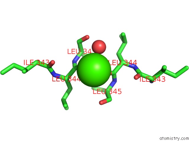
Mono view
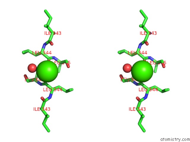
Stereo pair view
Mono view
Stereo pair view
A full contact list of Calcium with other atoms in the Ca binding
site number 1 of Crystal Structure of the Human RPAP3(TPR2)-PIH1D1(Cs) Complex within 5.0Å range:
|
Reference:
J.Henri,
M.E.Chagot,
M.Bourguet,
Y.Abel,
G.Terral,
C.Maurizy,
C.Aigueperse,
F.Georgescauld,
F.Vandermoere,
R.Saint-Fort,
I.Behm-Ansmant,
B.Charpentier,
B.Pradet-Balade,
C.Verheggen,
E.Bertrand,
P.Meyer,
S.Cianferani,
X.Manival,
M.Quinternet.
Deep Structural Analysis of RPAP3 and PIH1D1, Two Components of the HSP90 Co-Chaperone R2TP Complex. Structure V. 26 1196 2018.
ISSN: ISSN 1878-4186
PubMed: 30033218
DOI: 10.1016/J.STR.2018.06.002
Page generated: Tue Jul 16 08:12:41 2024
ISSN: ISSN 1878-4186
PubMed: 30033218
DOI: 10.1016/J.STR.2018.06.002
Last articles
Zn in 9JYWZn in 9IR4
Zn in 9IR3
Zn in 9GMX
Zn in 9GMW
Zn in 9JEJ
Zn in 9ERF
Zn in 9ERE
Zn in 9EGV
Zn in 9EGW