Calcium »
PDB 6guz-6hhz »
6hf4 »
Calcium in PDB 6hf4: The Structure of BOMAN26B, A GH26 Beta-Mannanase From Bacteroides Ovatus, Complexed with G1M4
Protein crystallography data
The structure of The Structure of BOMAN26B, A GH26 Beta-Mannanase From Bacteroides Ovatus, Complexed with G1M4, PDB code: 6hf4
was solved by
V.Bagenholm,
D.T.Logan,
H.Stalbrand,
with X-Ray Crystallography technique. A brief refinement statistics is given in the table below:
| Resolution Low / High (Å) | 47.50 / 1.78 |
| Space group | P 21 21 21 |
| Cell size a, b, c (Å), α, β, γ (°) | 50.122, 68.363, 94.993, 90.00, 90.00, 90.00 |
| R / Rfree (%) | 17.8 / 21 |
Other elements in 6hf4:
The structure of The Structure of BOMAN26B, A GH26 Beta-Mannanase From Bacteroides Ovatus, Complexed with G1M4 also contains other interesting chemical elements:
| Chlorine | (Cl) | 1 atom |
Calcium Binding Sites:
The binding sites of Calcium atom in the The Structure of BOMAN26B, A GH26 Beta-Mannanase From Bacteroides Ovatus, Complexed with G1M4
(pdb code 6hf4). This binding sites where shown within
5.0 Angstroms radius around Calcium atom.
In total only one binding site of Calcium was determined in the The Structure of BOMAN26B, A GH26 Beta-Mannanase From Bacteroides Ovatus, Complexed with G1M4, PDB code: 6hf4:
In total only one binding site of Calcium was determined in the The Structure of BOMAN26B, A GH26 Beta-Mannanase From Bacteroides Ovatus, Complexed with G1M4, PDB code: 6hf4:
Calcium binding site 1 out of 1 in 6hf4
Go back to
Calcium binding site 1 out
of 1 in the The Structure of BOMAN26B, A GH26 Beta-Mannanase From Bacteroides Ovatus, Complexed with G1M4
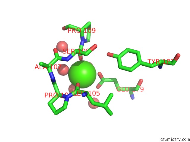
Mono view
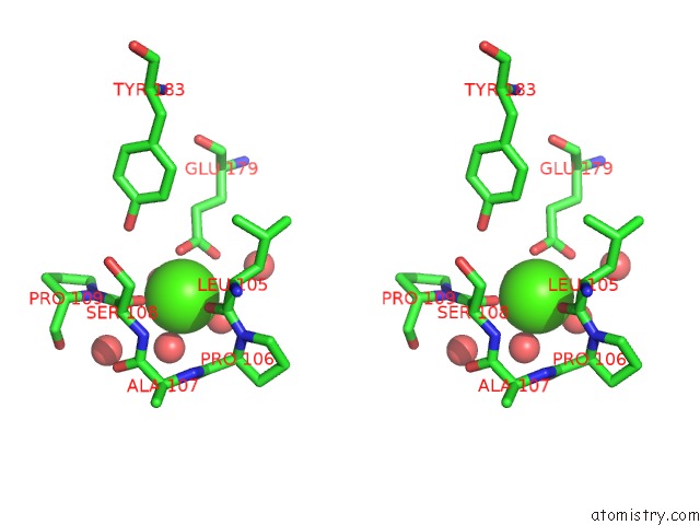
Stereo pair view
Mono view
Stereo pair view
A full contact list of Calcium with other atoms in the Ca binding
site number 1 of The Structure of BOMAN26B, A GH26 Beta-Mannanase From Bacteroides Ovatus, Complexed with G1M4 within 5.0Å range:
|
Reference:
V.Bagenholm,
M.Wiemann,
S.K.Reddy,
A.Bhattacharya,
A.Rosengren,
D.T.Logan,
H.Stalbrand.
A Surface-Exposed GH26 Beta-Mannanase Frombacteroides Ovatus: Structure, Role, and Phylogenetic Analysis OFBOMAN26B. J.Biol.Chem. V. 294 9100 2019.
ISSN: ESSN 1083-351X
PubMed: 31000630
DOI: 10.1074/JBC.RA118.007171
Page generated: Tue Jul 16 08:21:27 2024
ISSN: ESSN 1083-351X
PubMed: 31000630
DOI: 10.1074/JBC.RA118.007171
Last articles
Zn in 9J0NZn in 9J0O
Zn in 9J0P
Zn in 9FJX
Zn in 9EKB
Zn in 9C0F
Zn in 9CAH
Zn in 9CH0
Zn in 9CH3
Zn in 9CH1