Calcium »
PDB 6hi0-6i1q »
6hm3 »
Calcium in PDB 6hm3: Crystal Structure of RAD4 BRCT1,2 in Complex with A SLD3 Phosphopeptide
Protein crystallography data
The structure of Crystal Structure of RAD4 BRCT1,2 in Complex with A SLD3 Phosphopeptide, PDB code: 6hm3
was solved by
M.Day,
M.Rappas,
A.W.Oliver,
L.H.Pearl,
with X-Ray Crystallography technique. A brief refinement statistics is given in the table below:
| Resolution Low / High (Å) | 42.20 / 1.77 |
| Space group | P 21 21 21 |
| Cell size a, b, c (Å), α, β, γ (°) | 39.400, 59.170, 120.430, 90.00, 90.00, 90.00 |
| R / Rfree (%) | 19.2 / 22.8 |
Calcium Binding Sites:
The binding sites of Calcium atom in the Crystal Structure of RAD4 BRCT1,2 in Complex with A SLD3 Phosphopeptide
(pdb code 6hm3). This binding sites where shown within
5.0 Angstroms radius around Calcium atom.
In total 2 binding sites of Calcium where determined in the Crystal Structure of RAD4 BRCT1,2 in Complex with A SLD3 Phosphopeptide, PDB code: 6hm3:
Jump to Calcium binding site number: 1; 2;
In total 2 binding sites of Calcium where determined in the Crystal Structure of RAD4 BRCT1,2 in Complex with A SLD3 Phosphopeptide, PDB code: 6hm3:
Jump to Calcium binding site number: 1; 2;
Calcium binding site 1 out of 2 in 6hm3
Go back to
Calcium binding site 1 out
of 2 in the Crystal Structure of RAD4 BRCT1,2 in Complex with A SLD3 Phosphopeptide
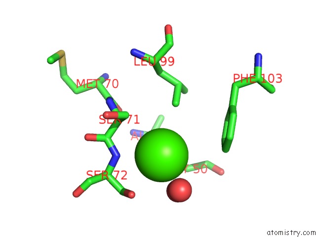
Mono view
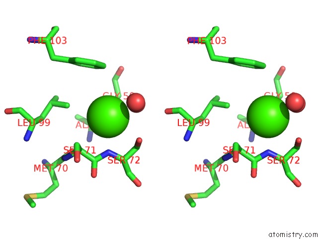
Stereo pair view
Mono view
Stereo pair view
A full contact list of Calcium with other atoms in the Ca binding
site number 1 of Crystal Structure of RAD4 BRCT1,2 in Complex with A SLD3 Phosphopeptide within 5.0Å range:
|
Calcium binding site 2 out of 2 in 6hm3
Go back to
Calcium binding site 2 out
of 2 in the Crystal Structure of RAD4 BRCT1,2 in Complex with A SLD3 Phosphopeptide
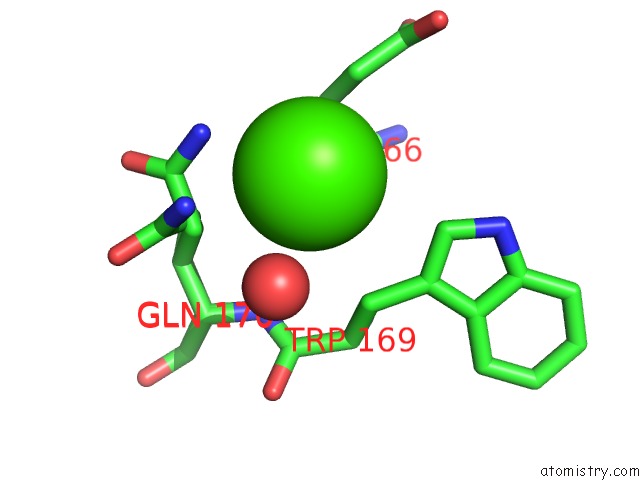
Mono view

Stereo pair view
Mono view
Stereo pair view
A full contact list of Calcium with other atoms in the Ca binding
site number 2 of Crystal Structure of RAD4 BRCT1,2 in Complex with A SLD3 Phosphopeptide within 5.0Å range:
|
Reference:
M.Day,
M.Rappas,
K.Ptasinska,
D.Boos,
A.W.Oliver,
L.H.Pearl.
Brct Domains of the Dna Damage Checkpoint Proteins TOPBP1/RAD4 Display Distinct Specificities For Phosphopeptide Ligands. Elife V. 7 2018.
ISSN: ESSN 2050-084X
PubMed: 30295604
DOI: 10.7554/ELIFE.39979
Page generated: Wed Jul 9 14:39:16 2025
ISSN: ESSN 2050-084X
PubMed: 30295604
DOI: 10.7554/ELIFE.39979
Last articles
Ca in 7KPJCa in 7KR5
Ca in 7KOQ
Ca in 7KLD
Ca in 7KOO
Ca in 7KNV
Ca in 7KLF
Ca in 7KLE
Ca in 7KMQ
Ca in 7KIQ