Calcium »
PDB 6hi0-6i1q »
6hos »
Calcium in PDB 6hos: Structure of the KPFLO2 Adhesin Domain in Complex with Glycerol
Protein crystallography data
The structure of Structure of the KPFLO2 Adhesin Domain in Complex with Glycerol, PDB code: 6hos
was solved by
L.-O.Essen,
M.Kock,
M.Veelders,
with X-Ray Crystallography technique. A brief refinement statistics is given in the table below:
| Resolution Low / High (Å) | 39.41 / 2.15 |
| Space group | C 1 2 1 |
| Cell size a, b, c (Å), α, β, γ (°) | 79.943, 103.186, 72.099, 90.00, 113.51, 90.00 |
| R / Rfree (%) | 17.2 / 19.9 |
Other elements in 6hos:
The structure of Structure of the KPFLO2 Adhesin Domain in Complex with Glycerol also contains other interesting chemical elements:
| Magnesium | (Mg) | 4 atoms |
Calcium Binding Sites:
The binding sites of Calcium atom in the Structure of the KPFLO2 Adhesin Domain in Complex with Glycerol
(pdb code 6hos). This binding sites where shown within
5.0 Angstroms radius around Calcium atom.
In total 2 binding sites of Calcium where determined in the Structure of the KPFLO2 Adhesin Domain in Complex with Glycerol, PDB code: 6hos:
Jump to Calcium binding site number: 1; 2;
In total 2 binding sites of Calcium where determined in the Structure of the KPFLO2 Adhesin Domain in Complex with Glycerol, PDB code: 6hos:
Jump to Calcium binding site number: 1; 2;
Calcium binding site 1 out of 2 in 6hos
Go back to
Calcium binding site 1 out
of 2 in the Structure of the KPFLO2 Adhesin Domain in Complex with Glycerol
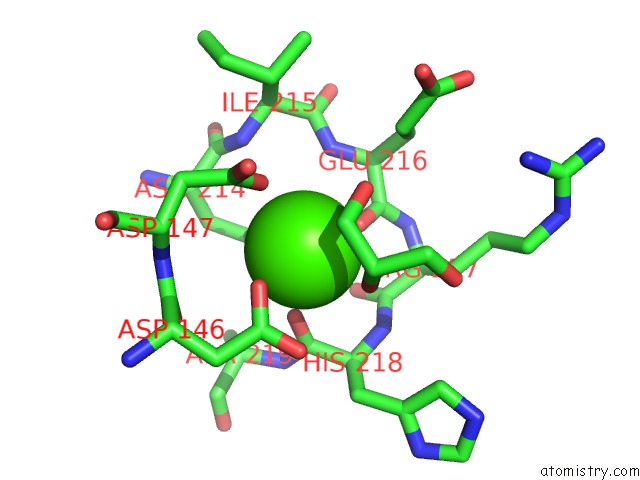
Mono view
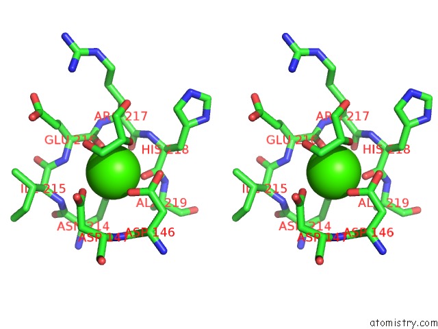
Stereo pair view
Mono view
Stereo pair view
A full contact list of Calcium with other atoms in the Ca binding
site number 1 of Structure of the KPFLO2 Adhesin Domain in Complex with Glycerol within 5.0Å range:
|
Calcium binding site 2 out of 2 in 6hos
Go back to
Calcium binding site 2 out
of 2 in the Structure of the KPFLO2 Adhesin Domain in Complex with Glycerol
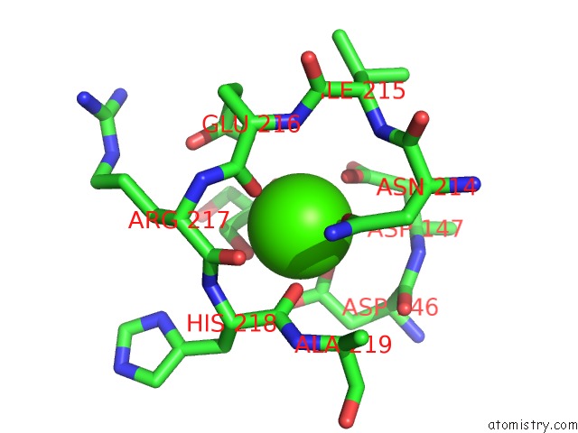
Mono view
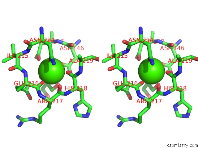
Stereo pair view
Mono view
Stereo pair view
A full contact list of Calcium with other atoms in the Ca binding
site number 2 of Structure of the KPFLO2 Adhesin Domain in Complex with Glycerol within 5.0Å range:
|
Reference:
M.Kock,
S.Bruckner,
N.Wozniak,
M.Maestre-Reyna,
M.Veelders,
J.Schlereth,
H.U.Mosch,
L.O.Essen.
Structural and Functional Characterization of PA14/FLO5-Like Adhesins Fromkomagataella Pastoris. Front Microbiol V. 9 2581 2018.
ISSN: ESSN 1664-302X
PubMed: 30425696
DOI: 10.3389/FMICB.2018.02581
Page generated: Tue Jul 16 08:28:50 2024
ISSN: ESSN 1664-302X
PubMed: 30425696
DOI: 10.3389/FMICB.2018.02581
Last articles
Zn in 9J0NZn in 9J0O
Zn in 9J0P
Zn in 9FJX
Zn in 9EKB
Zn in 9C0F
Zn in 9CAH
Zn in 9CH0
Zn in 9CH3
Zn in 9CH1