Calcium »
PDB 6hi0-6i1q »
6hq5 »
Calcium in PDB 6hq5: Structure of Eal Enzyme BD1971 - Camp and Cyclic-Di-Gmp Bound Form
Protein crystallography data
The structure of Structure of Eal Enzyme BD1971 - Camp and Cyclic-Di-Gmp Bound Form, PDB code: 6hq5
was solved by
A.L.Lovering,
I.T.Cadby,
with X-Ray Crystallography technique. A brief refinement statistics is given in the table below:
| Resolution Low / High (Å) | 64.31 / 2.83 |
| Space group | P 31 |
| Cell size a, b, c (Å), α, β, γ (°) | 83.857, 83.857, 138.454, 90.00, 90.00, 120.00 |
| R / Rfree (%) | 22.3 / 26.8 |
Calcium Binding Sites:
The binding sites of Calcium atom in the Structure of Eal Enzyme BD1971 - Camp and Cyclic-Di-Gmp Bound Form
(pdb code 6hq5). This binding sites where shown within
5.0 Angstroms radius around Calcium atom.
In total 4 binding sites of Calcium where determined in the Structure of Eal Enzyme BD1971 - Camp and Cyclic-Di-Gmp Bound Form, PDB code: 6hq5:
Jump to Calcium binding site number: 1; 2; 3; 4;
In total 4 binding sites of Calcium where determined in the Structure of Eal Enzyme BD1971 - Camp and Cyclic-Di-Gmp Bound Form, PDB code: 6hq5:
Jump to Calcium binding site number: 1; 2; 3; 4;
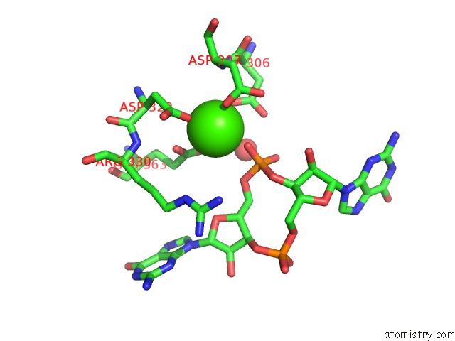
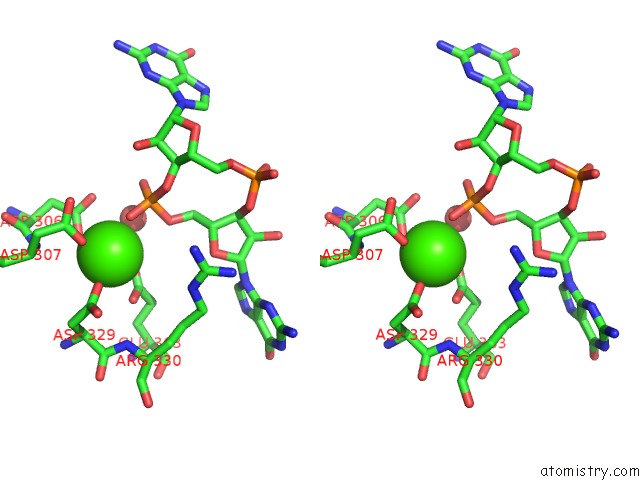
Calcium binding site 1 out of 4 in 6hq5
Go back to
Calcium binding site 1 out
of 4 in the Structure of Eal Enzyme BD1971 - Camp and Cyclic-Di-Gmp Bound Form
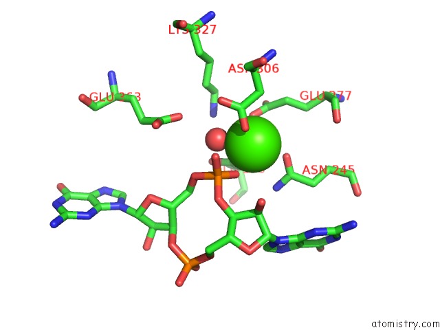
Mono view
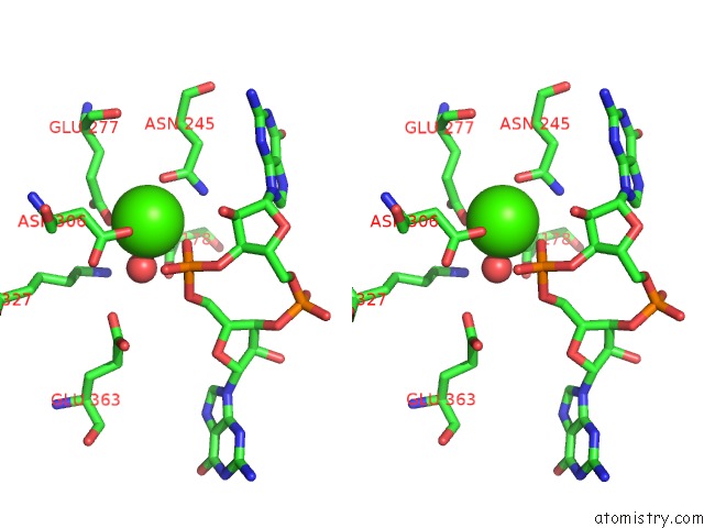
Stereo pair view
Mono view
Stereo pair view
A full contact list of Calcium with other atoms in the Ca binding
site number 1 of Structure of Eal Enzyme BD1971 - Camp and Cyclic-Di-Gmp Bound Form within 5.0Å range:
|
Calcium binding site 2 out of 4 in 6hq5
Go back to
Calcium binding site 2 out
of 4 in the Structure of Eal Enzyme BD1971 - Camp and Cyclic-Di-Gmp Bound Form
Mono view

Stereo pair view
Mono view
Stereo pair view
A full contact list of Calcium with other atoms in the Ca binding
site number 2 of Structure of Eal Enzyme BD1971 - Camp and Cyclic-Di-Gmp Bound Form within 5.0Å range:
|
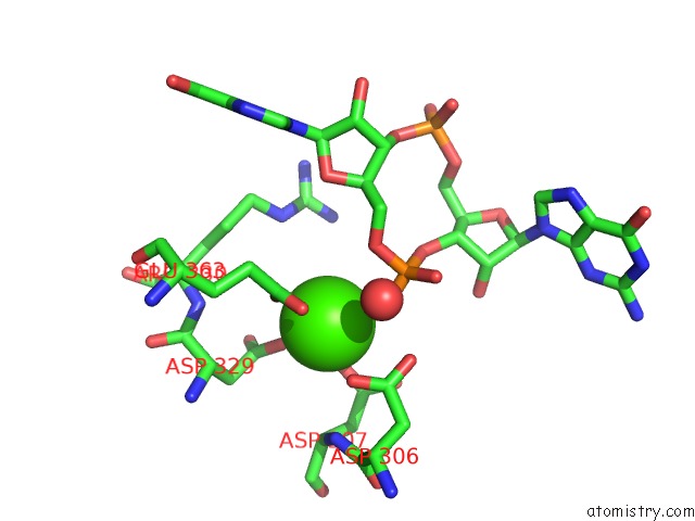
Calcium binding site 3 out of 4 in 6hq5
Go back to
Calcium binding site 3 out
of 4 in the Structure of Eal Enzyme BD1971 - Camp and Cyclic-Di-Gmp Bound Form
Mono view

Stereo pair view
Mono view
Stereo pair view
A full contact list of Calcium with other atoms in the Ca binding
site number 3 of Structure of Eal Enzyme BD1971 - Camp and Cyclic-Di-Gmp Bound Form within 5.0Å range:
|
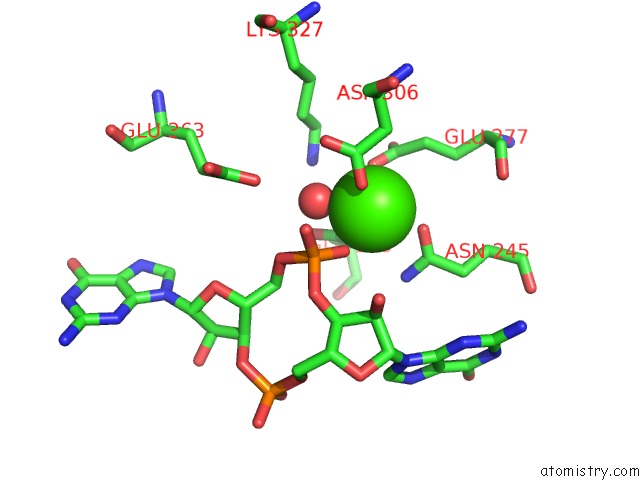
Calcium binding site 4 out of 4 in 6hq5
Go back to
Calcium binding site 4 out
of 4 in the Structure of Eal Enzyme BD1971 - Camp and Cyclic-Di-Gmp Bound Form
Mono view
Stereo pair view
Mono view
Stereo pair view
A full contact list of Calcium with other atoms in the Ca binding
site number 4 of Structure of Eal Enzyme BD1971 - Camp and Cyclic-Di-Gmp Bound Form within 5.0Å range:
|
Reference:
I.T.Cadby,
S.M.Basford,
R.Nottingham,
R.Meek,
R.Lowry,
C.Lambert,
M.Tridgett,
R.Till,
R.Ahmad,
R.Fung,
L.Hobley,
W.S.Hughes,
P.J.Moynihan,
R.E.Sockett,
A.L.Lovering.
Nucleotide Signaling Pathway Convergence in A Camp-Sensing Bacterial C-Di-Gmp Phosphodiesterase. Embo J. V. 38 00772 2019.
ISSN: ESSN 1460-2075
PubMed: 31355487
DOI: 10.15252/EMBJ.2018100772
Page generated: Wed Jul 9 14:40:54 2025
ISSN: ESSN 1460-2075
PubMed: 31355487
DOI: 10.15252/EMBJ.2018100772
Last articles
F in 7KZ4F in 7KYV
F in 7KYT
F in 7KYB
F in 7KYC
F in 7KYA
F in 7KY5
F in 7KXW
F in 7KY9
F in 7KWA