Calcium »
PDB 6hi0-6i1q »
6hqb »
Calcium in PDB 6hqb: Monomeric Cyanobacterial Photosystem I
Enzymatic activity of Monomeric Cyanobacterial Photosystem I
All present enzymatic activity of Monomeric Cyanobacterial Photosystem I:
1.97.1.12;
1.97.1.12;
Protein crystallography data
The structure of Monomeric Cyanobacterial Photosystem I, PDB code: 6hqb
was solved by
S.Y.Netzer-El,
N.Nelson,
I.Caspy,
with X-Ray Crystallography technique. A brief refinement statistics is given in the table below:
| Resolution Low / High (Å) | 49.29 / 4.00 |
| Space group | P 21 21 21 |
| Cell size a, b, c (Å), α, β, γ (°) | 124.322, 178.658, 181.446, 90.00, 90.00, 90.00 |
| R / Rfree (%) | 25.5 / 29.5 |
Other elements in 6hqb:
The structure of Monomeric Cyanobacterial Photosystem I also contains other interesting chemical elements:
| Magnesium | (Mg) | 93 atoms |
| Iron | (Fe) | 12 atoms |
| Chlorine | (Cl) | 1 atom |
Calcium Binding Sites:
The binding sites of Calcium atom in the Monomeric Cyanobacterial Photosystem I
(pdb code 6hqb). This binding sites where shown within
5.0 Angstroms radius around Calcium atom.
In total 2 binding sites of Calcium where determined in the Monomeric Cyanobacterial Photosystem I, PDB code: 6hqb:
Jump to Calcium binding site number: 1; 2;
In total 2 binding sites of Calcium where determined in the Monomeric Cyanobacterial Photosystem I, PDB code: 6hqb:
Jump to Calcium binding site number: 1; 2;
Calcium binding site 1 out of 2 in 6hqb
Go back to
Calcium binding site 1 out
of 2 in the Monomeric Cyanobacterial Photosystem I
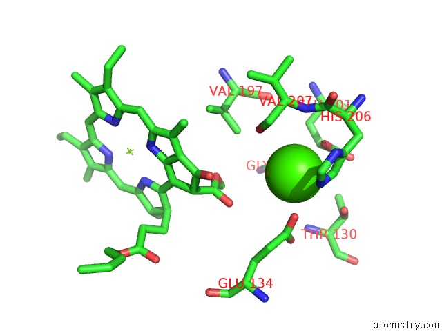
Mono view
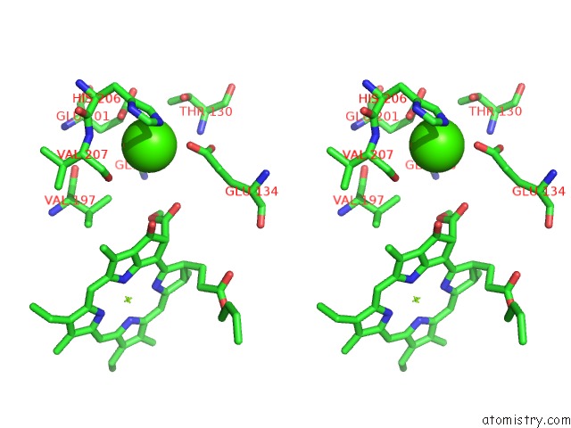
Stereo pair view
Mono view
Stereo pair view
A full contact list of Calcium with other atoms in the Ca binding
site number 1 of Monomeric Cyanobacterial Photosystem I within 5.0Å range:
|
Calcium binding site 2 out of 2 in 6hqb
Go back to
Calcium binding site 2 out
of 2 in the Monomeric Cyanobacterial Photosystem I
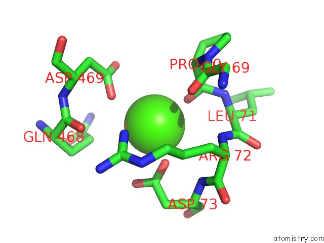
Mono view
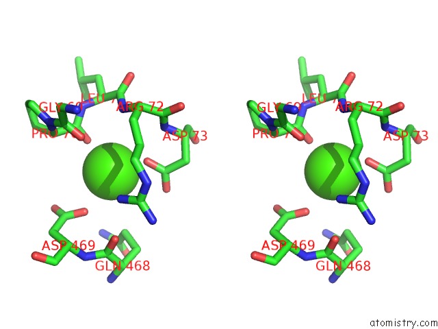
Stereo pair view
Mono view
Stereo pair view
A full contact list of Calcium with other atoms in the Ca binding
site number 2 of Monomeric Cyanobacterial Photosystem I within 5.0Å range:
|
Reference:
S.Y.Netzer-El,
I.Caspy,
N.Nelson.
Crystal Structure of Photosystem I Monomer From Synechocystis Pcc 6803. Front Plant Sci V. 9 1865 2018.
ISSN: ESSN 1664-462X
PubMed: 30662446
DOI: 10.3389/FPLS.2018.01865
Page generated: Wed Jul 9 14:41:57 2025
ISSN: ESSN 1664-462X
PubMed: 30662446
DOI: 10.3389/FPLS.2018.01865
Last articles
Cl in 5FZACl in 5FZ9
Cl in 5FZ8
Cl in 5FZ0
Cl in 5FZ7
Cl in 5FZ6
Cl in 5FZ1
Cl in 5FZ4
Cl in 5FZ3
Cl in 5FYY