Calcium »
PDB 6hhz-6i02 »
6hv2 »
Calcium in PDB 6hv2: Mmp-13 in Complex with the Peptide Imisf
Protein crystallography data
The structure of Mmp-13 in Complex with the Peptide Imisf, PDB code: 6hv2
was solved by
P.Mittl,
R.Riedl,
D.Hohl,
with X-Ray Crystallography technique. A brief refinement statistics is given in the table below:
| Resolution Low / High (Å) | 34.46 / 1.71 |
| Space group | P 61 2 2 |
| Cell size a, b, c (Å), α, β, γ (°) | 68.928, 68.928, 133.563, 90.00, 90.00, 120.00 |
| R / Rfree (%) | 21.4 / 26.3 |
Other elements in 6hv2:
The structure of Mmp-13 in Complex with the Peptide Imisf also contains other interesting chemical elements:
| Zinc | (Zn) | 2 atoms |
Calcium Binding Sites:
The binding sites of Calcium atom in the Mmp-13 in Complex with the Peptide Imisf
(pdb code 6hv2). This binding sites where shown within
5.0 Angstroms radius around Calcium atom.
In total 3 binding sites of Calcium where determined in the Mmp-13 in Complex with the Peptide Imisf, PDB code: 6hv2:
Jump to Calcium binding site number: 1; 2; 3;
In total 3 binding sites of Calcium where determined in the Mmp-13 in Complex with the Peptide Imisf, PDB code: 6hv2:
Jump to Calcium binding site number: 1; 2; 3;
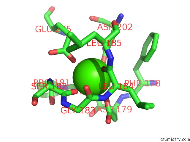
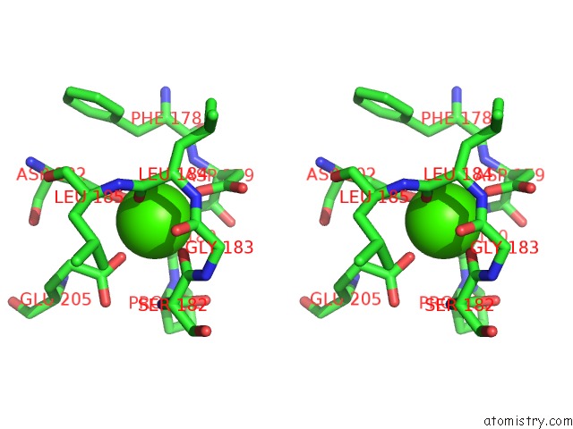
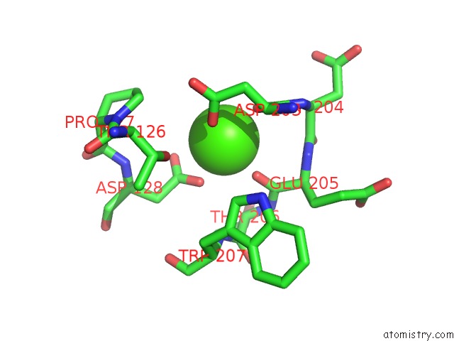
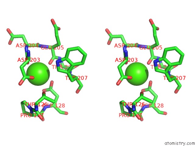
Calcium binding site 1 out of 3 in 6hv2
Go back to
Calcium binding site 1 out
of 3 in the Mmp-13 in Complex with the Peptide Imisf
Mono view
Stereo pair view
Mono view
Stereo pair view
A full contact list of Calcium with other atoms in the Ca binding
site number 1 of Mmp-13 in Complex with the Peptide Imisf within 5.0Å range:
|
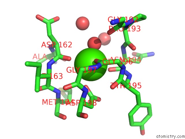
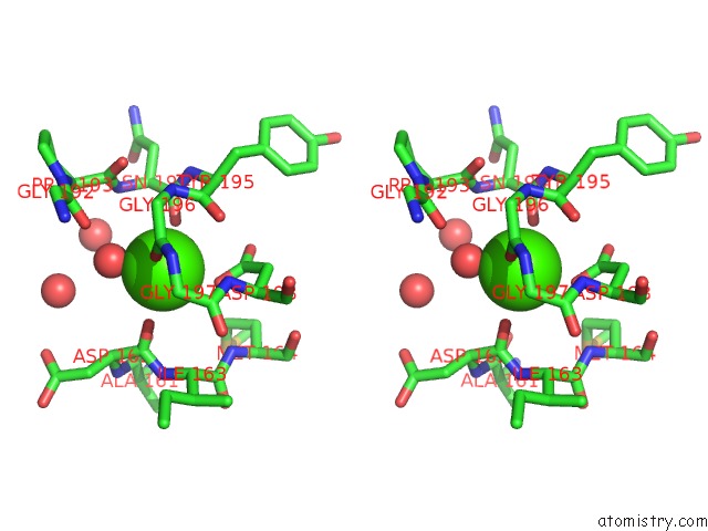
Calcium binding site 2 out of 3 in 6hv2
Go back to
Calcium binding site 2 out
of 3 in the Mmp-13 in Complex with the Peptide Imisf
Mono view
Stereo pair view
Mono view
Stereo pair view
A full contact list of Calcium with other atoms in the Ca binding
site number 2 of Mmp-13 in Complex with the Peptide Imisf within 5.0Å range:
|
Calcium binding site 3 out of 3 in 6hv2
Go back to
Calcium binding site 3 out
of 3 in the Mmp-13 in Complex with the Peptide Imisf
Mono view
Stereo pair view
Mono view
Stereo pair view
A full contact list of Calcium with other atoms in the Ca binding
site number 3 of Mmp-13 in Complex with the Peptide Imisf within 5.0Å range:
|
Reference:
F.M.Gall,
D.Hohl,
D.Frasson,
T.Wermelinger,
P.R.E.Mittl,
M.Sievers,
R.Riedl.
Drug Design Inspired By Nature: Crystallographic Detection of An Auto-Tailored Protease Inhibitor Template. Angew.Chem.Int.Ed.Engl. V. 58 4051 2019.
ISSN: ESSN 1521-3773
PubMed: 30615822
DOI: 10.1002/ANIE.201812348
Page generated: Tue Jul 16 08:32:41 2024
ISSN: ESSN 1521-3773
PubMed: 30615822
DOI: 10.1002/ANIE.201812348
Last articles
Zn in 9JYWZn in 9IR4
Zn in 9IR3
Zn in 9GMX
Zn in 9GMW
Zn in 9JEJ
Zn in 9ERF
Zn in 9ERE
Zn in 9EGV
Zn in 9EGW