Calcium »
PDB 6hi0-6i1q »
6hyj »
Calcium in PDB 6hyj: Psph Human Phosphoserine Phosphatase
Enzymatic activity of Psph Human Phosphoserine Phosphatase
All present enzymatic activity of Psph Human Phosphoserine Phosphatase:
3.1.3.3;
3.1.3.3;
Protein crystallography data
The structure of Psph Human Phosphoserine Phosphatase, PDB code: 6hyj
was solved by
J.Wouters,
M.Haufroid,
M.Mirgaux,
with X-Ray Crystallography technique. A brief refinement statistics is given in the table below:
| Resolution Low / High (Å) | 34.23 / 1.93 |
| Space group | C 2 2 21 |
| Cell size a, b, c (Å), α, β, γ (°) | 48.670, 128.970, 155.710, 90.00, 90.00, 90.00 |
| R / Rfree (%) | 20 / 26.3 |
Calcium Binding Sites:
The binding sites of Calcium atom in the Psph Human Phosphoserine Phosphatase
(pdb code 6hyj). This binding sites where shown within
5.0 Angstroms radius around Calcium atom.
In total 2 binding sites of Calcium where determined in the Psph Human Phosphoserine Phosphatase, PDB code: 6hyj:
Jump to Calcium binding site number: 1; 2;
In total 2 binding sites of Calcium where determined in the Psph Human Phosphoserine Phosphatase, PDB code: 6hyj:
Jump to Calcium binding site number: 1; 2;
Calcium binding site 1 out of 2 in 6hyj
Go back to
Calcium binding site 1 out
of 2 in the Psph Human Phosphoserine Phosphatase
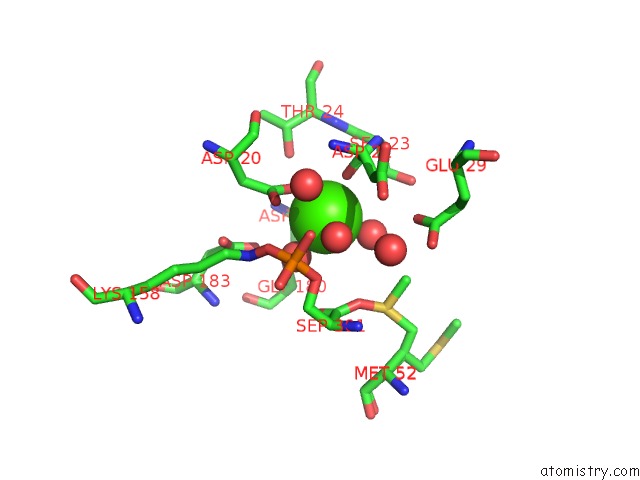
Mono view
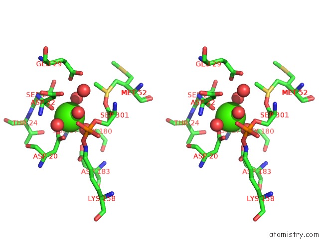
Stereo pair view
Mono view
Stereo pair view
A full contact list of Calcium with other atoms in the Ca binding
site number 1 of Psph Human Phosphoserine Phosphatase within 5.0Å range:
|
Calcium binding site 2 out of 2 in 6hyj
Go back to
Calcium binding site 2 out
of 2 in the Psph Human Phosphoserine Phosphatase
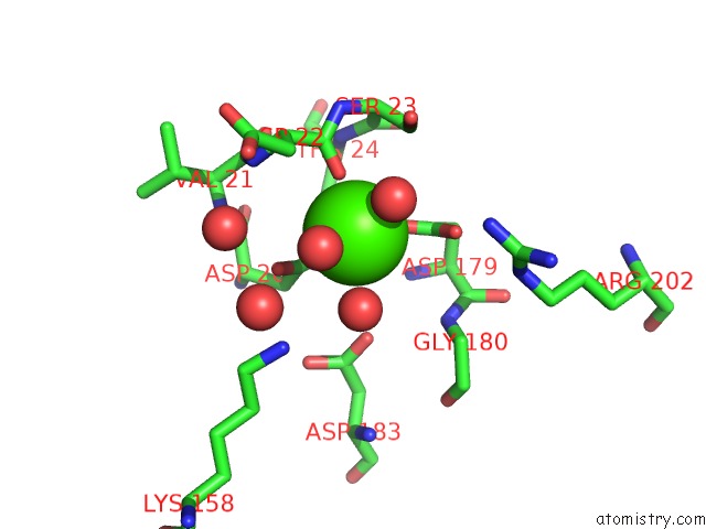
Mono view
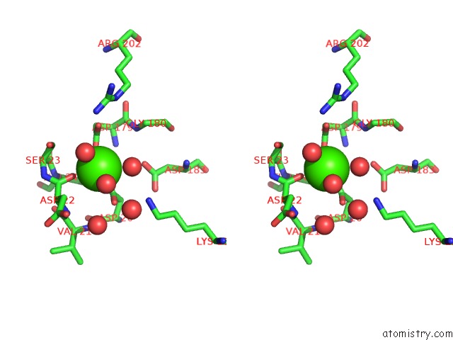
Stereo pair view
Mono view
Stereo pair view
A full contact list of Calcium with other atoms in the Ca binding
site number 2 of Psph Human Phosphoserine Phosphatase within 5.0Å range:
|
Reference:
M.Haufroid,
M.Mirgaux,
L.Leherte,
J.Wouters.
Crystal Structures and Snapshots Along the Reaction Pathway of Human Phosphoserine Phosphatase. Acta Crystallogr D Struct V. 75 592 2019BIOL.
ISSN: ISSN 2059-7983
PubMed: 31205021
DOI: 10.1107/S2059798319006867
Page generated: Tue Jul 16 08:34:49 2024
ISSN: ISSN 2059-7983
PubMed: 31205021
DOI: 10.1107/S2059798319006867
Last articles
Zn in 9MJ5Zn in 9HNW
Zn in 9G0L
Zn in 9FNE
Zn in 9DZN
Zn in 9E0I
Zn in 9D32
Zn in 9DAK
Zn in 8ZXC
Zn in 8ZUF