Calcium »
PDB 6hi0-6i1q »
6i02 »
Calcium in PDB 6i02: Structure of Human D-Glucuronyl C5 Epimerase in Complex with Product
Enzymatic activity of Structure of Human D-Glucuronyl C5 Epimerase in Complex with Product
All present enzymatic activity of Structure of Human D-Glucuronyl C5 Epimerase in Complex with Product:
5.1.3.17;
5.1.3.17;
Protein crystallography data
The structure of Structure of Human D-Glucuronyl C5 Epimerase in Complex with Product, PDB code: 6i02
was solved by
C.Debarnot,
Y.R.Monneau,
V.Roig-Zamboni,
C.Le Narvor,
A.Goulet,
F.Fadel,
R.R.Vives,
D.Bonnaffe,
H.Lortat-Jacob,
Y.Bourne,
with X-Ray Crystallography technique. A brief refinement statistics is given in the table below:
| Resolution Low / High (Å) | 49.06 / 2.45 |
| Space group | P 61 |
| Cell size a, b, c (Å), α, β, γ (°) | 99.811, 99.811, 260.488, 90.00, 90.00, 120.00 |
| R / Rfree (%) | 17.3 / 21.8 |
Calcium Binding Sites:
The binding sites of Calcium atom in the Structure of Human D-Glucuronyl C5 Epimerase in Complex with Product
(pdb code 6i02). This binding sites where shown within
5.0 Angstroms radius around Calcium atom.
In total 2 binding sites of Calcium where determined in the Structure of Human D-Glucuronyl C5 Epimerase in Complex with Product, PDB code: 6i02:
Jump to Calcium binding site number: 1; 2;
In total 2 binding sites of Calcium where determined in the Structure of Human D-Glucuronyl C5 Epimerase in Complex with Product, PDB code: 6i02:
Jump to Calcium binding site number: 1; 2;
Calcium binding site 1 out of 2 in 6i02
Go back to
Calcium binding site 1 out
of 2 in the Structure of Human D-Glucuronyl C5 Epimerase in Complex with Product
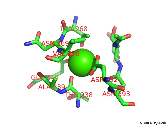
Mono view
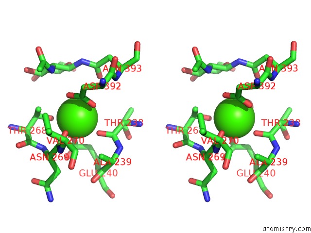
Stereo pair view
Mono view
Stereo pair view
A full contact list of Calcium with other atoms in the Ca binding
site number 1 of Structure of Human D-Glucuronyl C5 Epimerase in Complex with Product within 5.0Å range:
|
Calcium binding site 2 out of 2 in 6i02
Go back to
Calcium binding site 2 out
of 2 in the Structure of Human D-Glucuronyl C5 Epimerase in Complex with Product
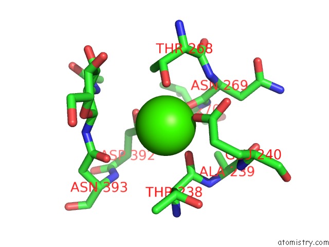
Mono view
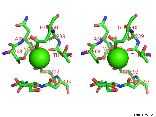
Stereo pair view
Mono view
Stereo pair view
A full contact list of Calcium with other atoms in the Ca binding
site number 2 of Structure of Human D-Glucuronyl C5 Epimerase in Complex with Product within 5.0Å range:
|
Reference:
C.Debarnot,
Y.R.Monneau,
V.Roig-Zamboni,
V.Delauzun,
C.Le Narvor,
E.Richard,
J.Henault,
A.Goulet,
F.Fadel,
R.R.Vives,
B.Priem,
D.Bonnaffe,
H.Lortat-Jacob,
Y.Bourne.
Substrate Binding Mode and Catalytic Mechanism of Human Heparan Sulfate D-Glucuronyl C5 Epimerase. Proc.Natl.Acad.Sci.Usa V. 116 6760 2019.
ISSN: ESSN 1091-6490
PubMed: 30872481
DOI: 10.1073/PNAS.1818333116
Page generated: Wed Jul 9 14:45:38 2025
ISSN: ESSN 1091-6490
PubMed: 30872481
DOI: 10.1073/PNAS.1818333116
Last articles
Ca in 7P9NCa in 7P9R
Ca in 7P8E
Ca in 7P7L
Ca in 7P7M
Ca in 7P7K
Ca in 7P7J
Ca in 7P73
Ca in 7P7E
Ca in 7P74