Calcium »
PDB 6i1t-6im1 »
6i4k »
Calcium in PDB 6i4k: Crystal Structure of Plasmodium Falciparum Actin I (G115A Mutant) in the Ca-Atp State
Protein crystallography data
The structure of Crystal Structure of Plasmodium Falciparum Actin I (G115A Mutant) in the Ca-Atp State, PDB code: 6i4k
was solved by
E.-P.Kumpula,
A.J.Lopez,
L.Tajedin,
H.Han,
I.Kursula,
with X-Ray Crystallography technique. A brief refinement statistics is given in the table below:
| Resolution Low / High (Å) | 43.80 / 1.83 |
| Space group | P 21 2 21 |
| Cell size a, b, c (Å), α, β, γ (°) | 69.830, 71.380, 110.930, 90.00, 90.00, 90.00 |
| R / Rfree (%) | 15.9 / 19.7 |
Other elements in 6i4k:
The structure of Crystal Structure of Plasmodium Falciparum Actin I (G115A Mutant) in the Ca-Atp State also contains other interesting chemical elements:
| Chlorine | (Cl) | 1 atom |
Calcium Binding Sites:
The binding sites of Calcium atom in the Crystal Structure of Plasmodium Falciparum Actin I (G115A Mutant) in the Ca-Atp State
(pdb code 6i4k). This binding sites where shown within
5.0 Angstroms radius around Calcium atom.
In total 3 binding sites of Calcium where determined in the Crystal Structure of Plasmodium Falciparum Actin I (G115A Mutant) in the Ca-Atp State, PDB code: 6i4k:
Jump to Calcium binding site number: 1; 2; 3;
In total 3 binding sites of Calcium where determined in the Crystal Structure of Plasmodium Falciparum Actin I (G115A Mutant) in the Ca-Atp State, PDB code: 6i4k:
Jump to Calcium binding site number: 1; 2; 3;
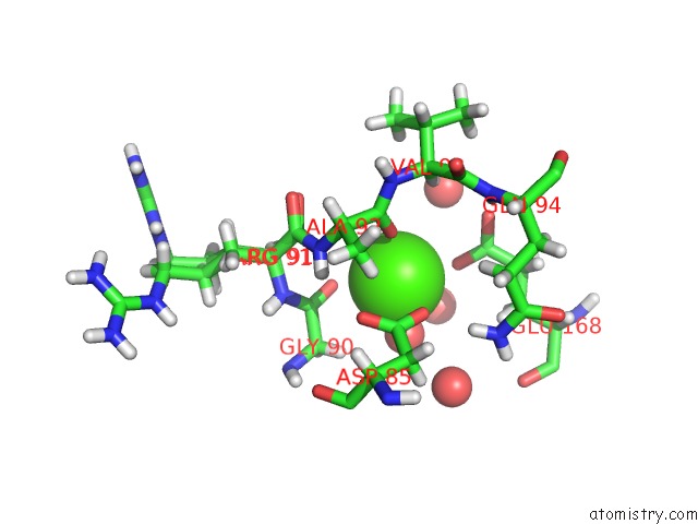
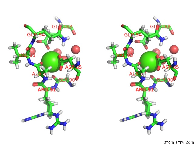
Calcium binding site 1 out of 3 in 6i4k
Go back to
Calcium binding site 1 out
of 3 in the Crystal Structure of Plasmodium Falciparum Actin I (G115A Mutant) in the Ca-Atp State
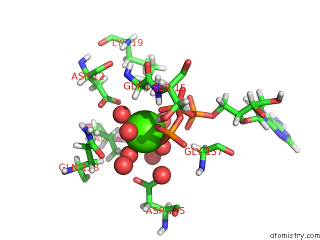
Mono view
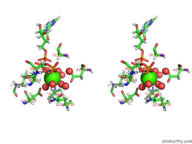
Stereo pair view
Mono view
Stereo pair view
A full contact list of Calcium with other atoms in the Ca binding
site number 1 of Crystal Structure of Plasmodium Falciparum Actin I (G115A Mutant) in the Ca-Atp State within 5.0Å range:
|
Calcium binding site 2 out of 3 in 6i4k
Go back to
Calcium binding site 2 out
of 3 in the Crystal Structure of Plasmodium Falciparum Actin I (G115A Mutant) in the Ca-Atp State
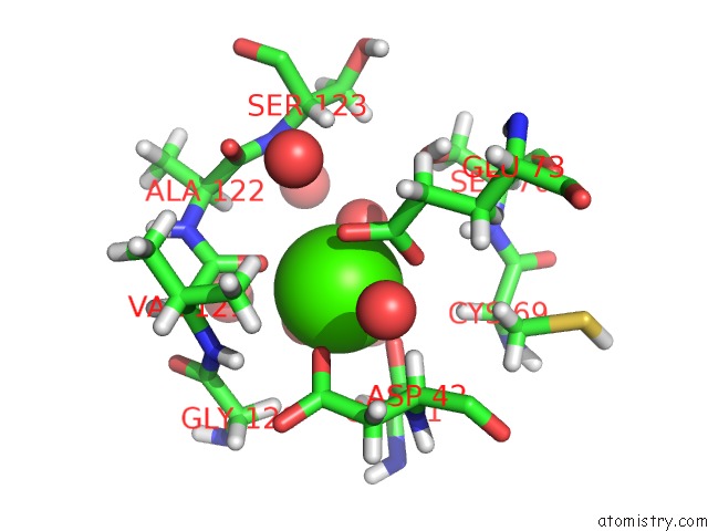
Mono view
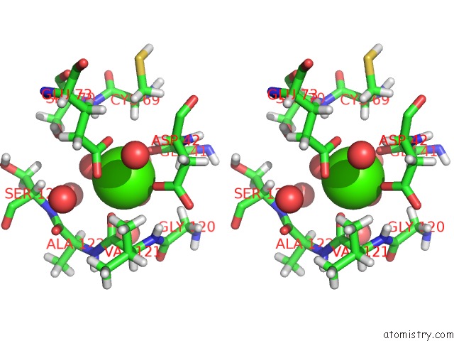
Stereo pair view
Mono view
Stereo pair view
A full contact list of Calcium with other atoms in the Ca binding
site number 2 of Crystal Structure of Plasmodium Falciparum Actin I (G115A Mutant) in the Ca-Atp State within 5.0Å range:
|
Calcium binding site 3 out of 3 in 6i4k
Go back to
Calcium binding site 3 out
of 3 in the Crystal Structure of Plasmodium Falciparum Actin I (G115A Mutant) in the Ca-Atp State
Mono view
Stereo pair view
Mono view
Stereo pair view
A full contact list of Calcium with other atoms in the Ca binding
site number 3 of Crystal Structure of Plasmodium Falciparum Actin I (G115A Mutant) in the Ca-Atp State within 5.0Å range:
|
Reference:
E.P.Kumpula,
A.J.Lopez,
L.Tajedin,
H.Han,
I.Kursula.
Atomic View Into Plasmodium Actin Polymerization, Atp Hydrolysis, and Fragmentation. Plos Biol. V. 17 00315 2019.
ISSN: ESSN 1545-7885
PubMed: 31199804
DOI: 10.1371/JOURNAL.PBIO.3000315
Page generated: Tue Jul 16 09:19:06 2024
ISSN: ESSN 1545-7885
PubMed: 31199804
DOI: 10.1371/JOURNAL.PBIO.3000315
Last articles
Zn in 9J0NZn in 9J0O
Zn in 9J0P
Zn in 9FJX
Zn in 9EKB
Zn in 9C0F
Zn in 9CAH
Zn in 9CH0
Zn in 9CH3
Zn in 9CH1