Calcium in PDB 6i4l: Crystal Structure of Plasmodium Falciparum Actin I (G115A Mutant) in the Mg-K-Atp/Adp State
Protein crystallography data
The structure of Crystal Structure of Plasmodium Falciparum Actin I (G115A Mutant) in the Mg-K-Atp/Adp State, PDB code: 6i4l
was solved by
E.-P.Kumpula,
A.J.Lopez,
L.Tajedin,
H.Han,
I.Kursula,
with X-Ray Crystallography technique. A brief refinement statistics is given in the table below:
Resolution Low / High (Å)
|
45.36 /
1.83
|
Space group
|
P 21 2 21
|
Cell size a, b, c (Å), α, β, γ (°)
|
69.450,
71.330,
110.310,
90.00,
90.00,
90.00
|
R / Rfree (%)
|
20 /
24.3
|
Other elements in 6i4l:
The structure of Crystal Structure of Plasmodium Falciparum Actin I (G115A Mutant) in the Mg-K-Atp/Adp State also contains other interesting chemical elements:
Calcium Binding Sites:
The binding sites of Calcium atom in the Crystal Structure of Plasmodium Falciparum Actin I (G115A Mutant) in the Mg-K-Atp/Adp State
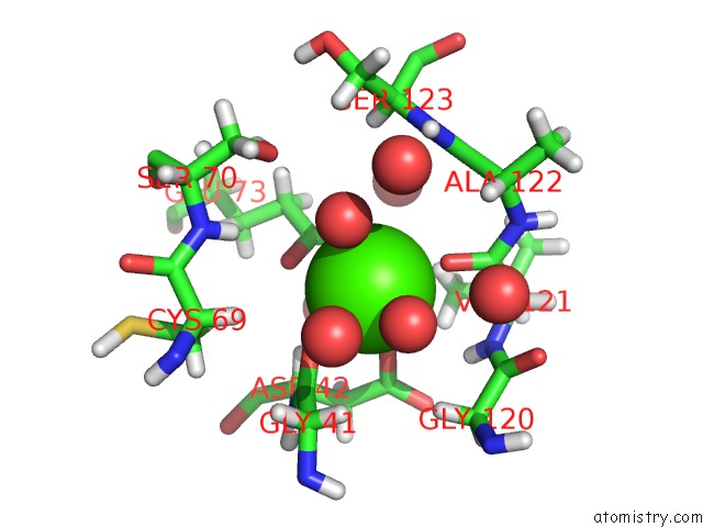
(pdb code 6i4l). This binding sites where shown within
5.0 Angstroms radius around Calcium atom.
In total 2 binding sites of Calcium where determined in the
Crystal Structure of Plasmodium Falciparum Actin I (G115A Mutant) in the Mg-K-Atp/Adp State, PDB code: 6i4l:
Jump to Calcium binding site number:
1;
2;
Calcium binding site 1 out
of 2 in 6i4l
Go back to
Calcium Binding Sites List in 6i4l
Calcium binding site 1 out
of 2 in the Crystal Structure of Plasmodium Falciparum Actin I (G115A Mutant) in the Mg-K-Atp/Adp State
Mono view
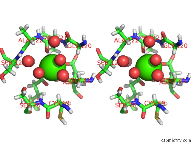
Stereo pair view
|
A full contact list of Calcium with other atoms in the Ca binding
site number 1 of Crystal Structure of Plasmodium Falciparum Actin I (G115A Mutant) in the Mg-K-Atp/Adp State within 5.0Å range:
probe
|
atom
|
residue
|
distance (Å)
|
B
|
Occ
|
G:Ca201
b:20.4
occ:1.00
|
OD1
|
G:ASP42
|
2.3
|
17.9
|
1.0
|
O
|
G:GLY41
|
2.3
|
18.7
|
1.0
|
O
|
G:VAL121
|
2.3
|
22.4
|
1.0
|
O
|
G:HOH381
|
2.4
|
23.5
|
1.0
|
OE2
|
G:GLU73
|
2.4
|
18.8
|
1.0
|
O
|
G:HOH377
|
2.5
|
17.0
|
1.0
|
OE1
|
G:GLU73
|
2.6
|
19.5
|
1.0
|
CD
|
G:GLU73
|
2.9
|
17.1
|
1.0
|
HA
|
G:ASP42
|
3.2
|
20.9
|
1.0
|
C
|
G:GLY41
|
3.3
|
20.0
|
1.0
|
HG13
|
G:VAL121
|
3.5
|
27.1
|
1.0
|
CG
|
G:ASP42
|
3.5
|
20.4
|
1.0
|
C
|
G:VAL121
|
3.5
|
22.1
|
1.0
|
H
|
G:VAL121
|
3.8
|
24.5
|
1.0
|
HA3
|
G:GLY120
|
3.9
|
30.4
|
1.0
|
N
|
G:VAL121
|
3.9
|
20.4
|
1.0
|
CA
|
G:ASP42
|
3.9
|
17.4
|
1.0
|
HA
|
G:CYS69
|
3.9
|
22.3
|
1.0
|
HG12
|
G:VAL121
|
4.0
|
27.1
|
1.0
|
N
|
G:ASP42
|
4.0
|
17.9
|
1.0
|
HA3
|
G:GLY41
|
4.1
|
25.6
|
1.0
|
H
|
G:SER70
|
4.1
|
21.4
|
1.0
|
CG1
|
G:VAL121
|
4.2
|
22.6
|
1.0
|
O
|
G:HOH339
|
4.2
|
15.5
|
1.0
|
CA
|
G:VAL121
|
4.3
|
21.5
|
1.0
|
CB
|
G:ASP42
|
4.3
|
18.1
|
1.0
|
HA
|
G:ALA122
|
4.3
|
28.8
|
1.0
|
C
|
G:GLY120
|
4.3
|
22.9
|
1.0
|
CA
|
G:GLY41
|
4.3
|
21.3
|
1.0
|
CG
|
G:GLU73
|
4.4
|
15.1
|
1.0
|
OD2
|
G:ASP42
|
4.4
|
21.7
|
1.0
|
N
|
G:ALA122
|
4.5
|
24.3
|
1.0
|
HB2
|
G:CYS69
|
4.6
|
22.9
|
1.0
|
C
|
G:ALA122
|
4.6
|
23.5
|
1.0
|
CA
|
G:GLY120
|
4.6
|
25.4
|
1.0
|
O
|
G:HOH383
|
4.6
|
22.7
|
1.0
|
O
|
G:HOH350
|
4.6
|
29.6
|
1.0
|
O
|
G:HOH384
|
4.6
|
27.2
|
1.0
|
OG
|
G:SER70
|
4.6
|
21.1
|
1.0
|
N
|
G:SER123
|
4.7
|
24.0
|
1.0
|
CA
|
G:ALA122
|
4.7
|
24.0
|
1.0
|
HA2
|
G:GLY41
|
4.7
|
25.6
|
1.0
|
HG3
|
G:GLU73
|
4.7
|
18.1
|
1.0
|
HB3
|
G:ASP42
|
4.7
|
21.8
|
1.0
|
O
|
G:HOH325
|
4.7
|
21.6
|
1.0
|
H
|
G:SER123
|
4.8
|
28.8
|
1.0
|
HG2
|
G:GLU73
|
4.8
|
18.1
|
1.0
|
HA
|
G:SER123
|
4.8
|
29.8
|
1.0
|
CA
|
G:CYS69
|
4.8
|
18.6
|
1.0
|
H
|
G:ASP42
|
4.9
|
21.5
|
1.0
|
CB
|
G:VAL121
|
4.9
|
25.4
|
1.0
|
O
|
G:ALA122
|
4.9
|
20.4
|
1.0
|
N
|
G:SER70
|
4.9
|
17.8
|
1.0
|
HG11
|
G:VAL121
|
4.9
|
27.1
|
1.0
|
O
|
G:GLY120
|
4.9
|
26.5
|
1.0
|
|
Calcium binding site 2 out
of 2 in 6i4l
Go back to
Calcium Binding Sites List in 6i4l
Calcium binding site 2 out
of 2 in the Crystal Structure of Plasmodium Falciparum Actin I (G115A Mutant) in the Mg-K-Atp/Adp State
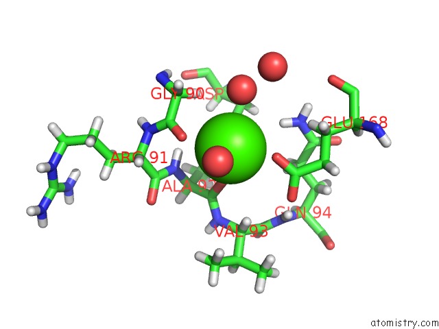
Mono view
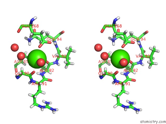
Stereo pair view
|
A full contact list of Calcium with other atoms in the Ca binding
site number 2 of Crystal Structure of Plasmodium Falciparum Actin I (G115A Mutant) in the Mg-K-Atp/Adp State within 5.0Å range:
probe
|
atom
|
residue
|
distance (Å)
|
B
|
Occ
|
G:Ca202
b:29.6
occ:1.00
|
O
|
G:ALA92
|
2.3
|
27.4
|
1.0
|
OD2
|
G:ASP85
|
2.4
|
26.4
|
1.0
|
O
|
G:HOH340
|
2.4
|
35.5
|
1.0
|
OE1
|
A:GLU168
|
2.5
|
24.0
|
1.0
|
O
|
G:GLY90
|
2.6
|
27.9
|
1.0
|
O
|
G:HOH365
|
3.0
|
29.7
|
1.0
|
OD1
|
G:ASP85
|
3.0
|
23.1
|
1.0
|
CG
|
G:ASP85
|
3.0
|
22.9
|
1.0
|
C
|
G:GLY90
|
3.4
|
27.6
|
1.0
|
HA2
|
G:GLY90
|
3.4
|
29.2
|
1.0
|
CD
|
A:GLU168
|
3.5
|
23.7
|
1.0
|
C
|
G:ALA92
|
3.5
|
26.0
|
1.0
|
H
|
G:ALA92
|
3.7
|
29.2
|
1.0
|
HE21
|
G:GLN94
|
3.8
|
23.5
|
1.0
|
CA
|
G:GLY90
|
3.9
|
24.3
|
1.0
|
N
|
G:ALA92
|
3.9
|
24.3
|
1.0
|
OE2
|
A:GLU168
|
3.9
|
23.9
|
1.0
|
HG3
|
G:GLN94
|
4.0
|
21.9
|
1.0
|
HA
|
G:VAL93
|
4.0
|
31.8
|
1.0
|
HA3
|
G:GLY90
|
4.1
|
29.2
|
1.0
|
HB3
|
A:GLU168
|
4.3
|
24.1
|
1.0
|
CA
|
G:ALA92
|
4.3
|
23.8
|
1.0
|
N
|
G:ARG91
|
4.3
|
27.3
|
1.0
|
HB3
|
G:ALA92
|
4.4
|
31.1
|
1.0
|
C
|
G:ARG91
|
4.4
|
26.6
|
1.0
|
CB
|
G:ASP85
|
4.5
|
22.5
|
1.0
|
NE2
|
G:GLN94
|
4.5
|
19.6
|
1.0
|
N
|
G:VAL93
|
4.6
|
24.8
|
1.0
|
HA
|
G:ARG91
|
4.6
|
37.2
|
1.0
|
HB3
|
G:ASP85
|
4.7
|
27.0
|
1.0
|
CA
|
G:ARG91
|
4.7
|
31.0
|
1.0
|
CG
|
A:GLU168
|
4.7
|
20.3
|
1.0
|
CA
|
G:VAL93
|
4.7
|
26.5
|
1.0
|
O
|
G:HOH348
|
4.8
|
31.5
|
1.0
|
CB
|
A:GLU168
|
4.8
|
20.1
|
1.0
|
CG
|
G:GLN94
|
4.8
|
18.3
|
1.0
|
HB2
|
G:ASP85
|
4.9
|
27.0
|
1.0
|
H
|
G:GLN94
|
4.9
|
27.1
|
1.0
|
CB
|
G:ALA92
|
4.9
|
26.0
|
1.0
|
HB2
|
A:GLU168
|
4.9
|
24.1
|
1.0
|
H
|
G:ARG91
|
5.0
|
32.7
|
1.0
|
|
Reference:
E.P.Kumpula,
A.J.Lopez,
L.Tajedin,
H.Han,
I.Kursula.
Atomic View Into Plasmodium Actin Polymerization, Atp Hydrolysis, and Fragmentation. Plos Biol. V. 17 00315 2019.
ISSN: ESSN 1545-7885
PubMed: 31199804
DOI: 10.1371/JOURNAL.PBIO.3000315
Page generated: Tue Jul 16 09:19:28 2024
|