Calcium »
PDB 6i1t-6im1 »
6i8a »
Calcium in PDB 6i8a: The Crystal Structure of the POL2 Catalytic Domain of Dna Polymerase Epsilon Carrying A P301R Substitution.
Enzymatic activity of The Crystal Structure of the POL2 Catalytic Domain of Dna Polymerase Epsilon Carrying A P301R Substitution.
All present enzymatic activity of The Crystal Structure of the POL2 Catalytic Domain of Dna Polymerase Epsilon Carrying A P301R Substitution.:
2.7.7.7;
2.7.7.7;
Protein crystallography data
The structure of The Crystal Structure of the POL2 Catalytic Domain of Dna Polymerase Epsilon Carrying A P301R Substitution., PDB code: 6i8a
was solved by
V.Parkash,
E.Johansson,
with X-Ray Crystallography technique. A brief refinement statistics is given in the table below:
| Resolution Low / High (Å) | 19.96 / 2.65 |
| Space group | P 1 2 1 |
| Cell size a, b, c (Å), α, β, γ (°) | 154.466, 70.257, 159.335, 90.00, 112.85, 90.00 |
| R / Rfree (%) | 22.9 / 27.9 |
Other elements in 6i8a:
The structure of The Crystal Structure of the POL2 Catalytic Domain of Dna Polymerase Epsilon Carrying A P301R Substitution. also contains other interesting chemical elements:
| Iron | (Fe) | 2 atoms |
Calcium Binding Sites:
The binding sites of Calcium atom in the The Crystal Structure of the POL2 Catalytic Domain of Dna Polymerase Epsilon Carrying A P301R Substitution.
(pdb code 6i8a). This binding sites where shown within
5.0 Angstroms radius around Calcium atom.
In total 4 binding sites of Calcium where determined in the The Crystal Structure of the POL2 Catalytic Domain of Dna Polymerase Epsilon Carrying A P301R Substitution., PDB code: 6i8a:
Jump to Calcium binding site number: 1; 2; 3; 4;
In total 4 binding sites of Calcium where determined in the The Crystal Structure of the POL2 Catalytic Domain of Dna Polymerase Epsilon Carrying A P301R Substitution., PDB code: 6i8a:
Jump to Calcium binding site number: 1; 2; 3; 4;
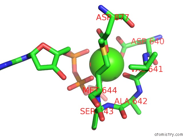
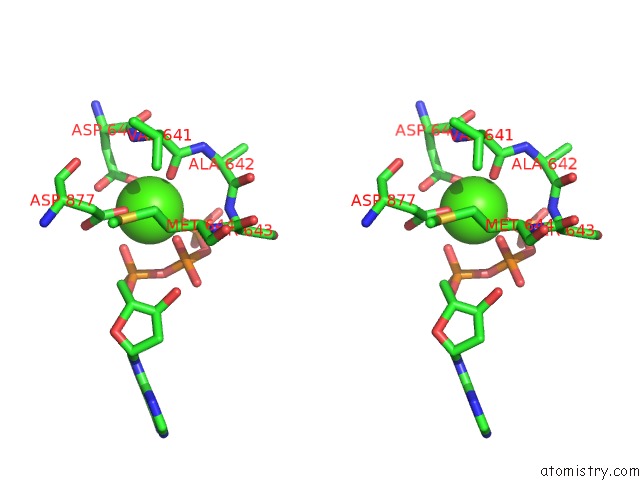
Calcium binding site 1 out of 4 in 6i8a
Go back to
Calcium binding site 1 out
of 4 in the The Crystal Structure of the POL2 Catalytic Domain of Dna Polymerase Epsilon Carrying A P301R Substitution.
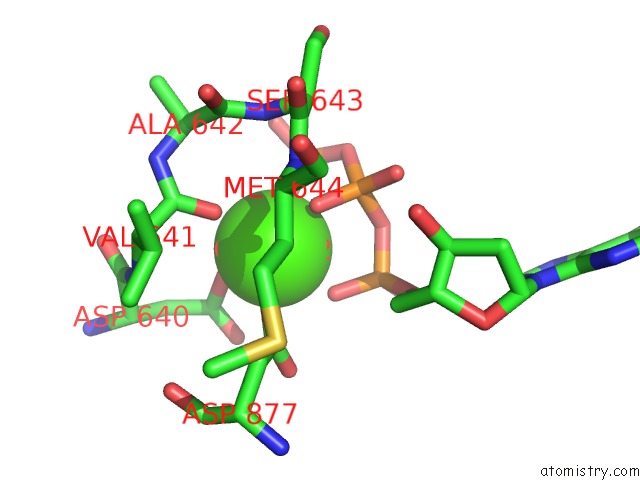
Mono view
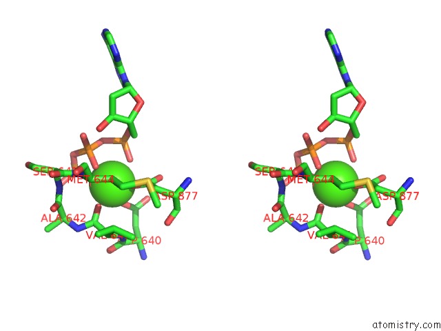
Stereo pair view
Mono view
Stereo pair view
A full contact list of Calcium with other atoms in the Ca binding
site number 1 of The Crystal Structure of the POL2 Catalytic Domain of Dna Polymerase Epsilon Carrying A P301R Substitution. within 5.0Å range:
|
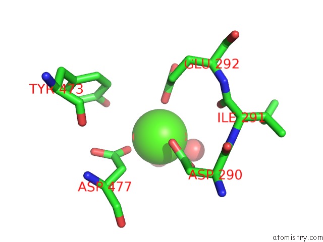
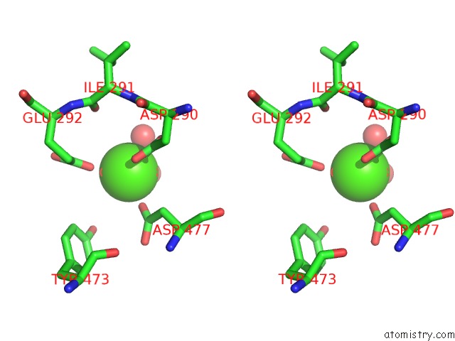
Calcium binding site 2 out of 4 in 6i8a
Go back to
Calcium binding site 2 out
of 4 in the The Crystal Structure of the POL2 Catalytic Domain of Dna Polymerase Epsilon Carrying A P301R Substitution.
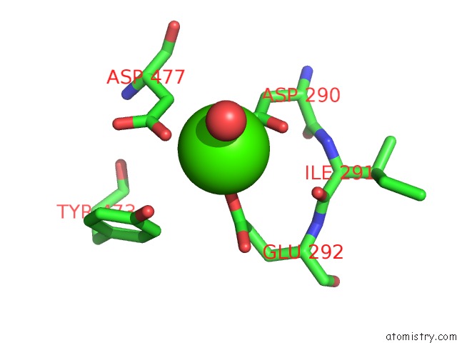
Mono view
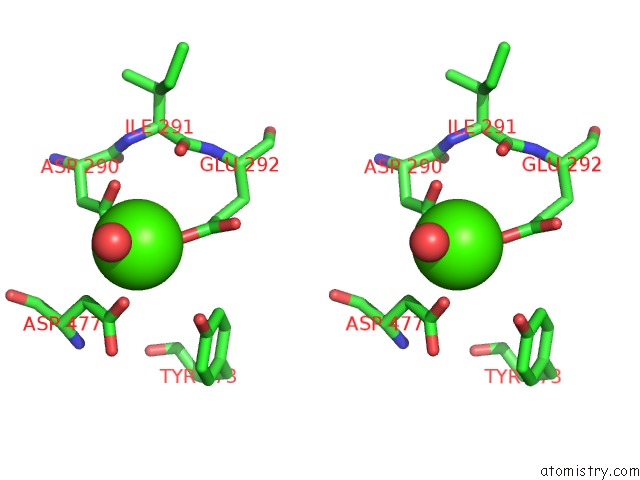
Stereo pair view
Mono view
Stereo pair view
A full contact list of Calcium with other atoms in the Ca binding
site number 2 of The Crystal Structure of the POL2 Catalytic Domain of Dna Polymerase Epsilon Carrying A P301R Substitution. within 5.0Å range:
|
Calcium binding site 3 out of 4 in 6i8a
Go back to
Calcium binding site 3 out
of 4 in the The Crystal Structure of the POL2 Catalytic Domain of Dna Polymerase Epsilon Carrying A P301R Substitution.
Mono view
Stereo pair view
Mono view
Stereo pair view
A full contact list of Calcium with other atoms in the Ca binding
site number 3 of The Crystal Structure of the POL2 Catalytic Domain of Dna Polymerase Epsilon Carrying A P301R Substitution. within 5.0Å range:
|
Calcium binding site 4 out of 4 in 6i8a
Go back to
Calcium binding site 4 out
of 4 in the The Crystal Structure of the POL2 Catalytic Domain of Dna Polymerase Epsilon Carrying A P301R Substitution.
Mono view
Stereo pair view
Mono view
Stereo pair view
A full contact list of Calcium with other atoms in the Ca binding
site number 4 of The Crystal Structure of the POL2 Catalytic Domain of Dna Polymerase Epsilon Carrying A P301R Substitution. within 5.0Å range:
|
Reference:
V.Parkash,
Y.Kulkarni,
J.Ter Beek,
P.V.Shcherbakova,
S.C.L.Kamerlin,
E.Johansson.
Structural Consequence of the Most Frequently Recurring Cancer-Associated Substitution in Dna Polymerase Epsilon. Nat Commun V. 10 373 2019.
ISSN: ESSN 2041-1723
PubMed: 30670696
DOI: 10.1038/S41467-018-08114-9
Page generated: Tue Jul 16 09:20:50 2024
ISSN: ESSN 2041-1723
PubMed: 30670696
DOI: 10.1038/S41467-018-08114-9
Last articles
Ca in 3I5LCa in 3I6N
Ca in 3I77
Ca in 3I5E
Ca in 3I5I
Ca in 3I5H
Ca in 3I5G
Ca in 3I57
Ca in 3I08
Ca in 3I46