Calcium »
PDB 6i1t-6im1 »
6ifi »
Calcium in PDB 6ifi: Crystal Structure of the Apo Form of Cmp-N-Acetylneuraminate Synthetase From Vibrio Cholerae
Enzymatic activity of Crystal Structure of the Apo Form of Cmp-N-Acetylneuraminate Synthetase From Vibrio Cholerae
All present enzymatic activity of Crystal Structure of the Apo Form of Cmp-N-Acetylneuraminate Synthetase From Vibrio Cholerae:
2.7.7.43;
2.7.7.43;
Protein crystallography data
The structure of Crystal Structure of the Apo Form of Cmp-N-Acetylneuraminate Synthetase From Vibrio Cholerae, PDB code: 6ifi
was solved by
S.Bose,
R.Subramanian,
with X-Ray Crystallography technique. A brief refinement statistics is given in the table below:
| Resolution Low / High (Å) | 25.08 / 2.80 |
| Space group | P 41 |
| Cell size a, b, c (Å), α, β, γ (°) | 75.250, 75.250, 109.590, 90.00, 90.00, 90.00 |
| R / Rfree (%) | 21.2 / 24 |
Calcium Binding Sites:
The binding sites of Calcium atom in the Crystal Structure of the Apo Form of Cmp-N-Acetylneuraminate Synthetase From Vibrio Cholerae
(pdb code 6ifi). This binding sites where shown within
5.0 Angstroms radius around Calcium atom.
In total 2 binding sites of Calcium where determined in the Crystal Structure of the Apo Form of Cmp-N-Acetylneuraminate Synthetase From Vibrio Cholerae, PDB code: 6ifi:
Jump to Calcium binding site number: 1; 2;
In total 2 binding sites of Calcium where determined in the Crystal Structure of the Apo Form of Cmp-N-Acetylneuraminate Synthetase From Vibrio Cholerae, PDB code: 6ifi:
Jump to Calcium binding site number: 1; 2;
Calcium binding site 1 out of 2 in 6ifi
Go back to
Calcium binding site 1 out
of 2 in the Crystal Structure of the Apo Form of Cmp-N-Acetylneuraminate Synthetase From Vibrio Cholerae
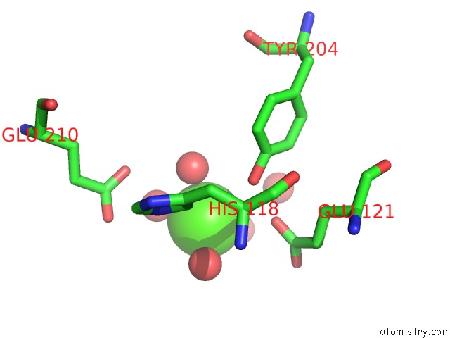
Mono view
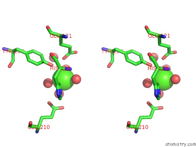
Stereo pair view
Mono view
Stereo pair view
A full contact list of Calcium with other atoms in the Ca binding
site number 1 of Crystal Structure of the Apo Form of Cmp-N-Acetylneuraminate Synthetase From Vibrio Cholerae within 5.0Å range:
|
Calcium binding site 2 out of 2 in 6ifi
Go back to
Calcium binding site 2 out
of 2 in the Crystal Structure of the Apo Form of Cmp-N-Acetylneuraminate Synthetase From Vibrio Cholerae
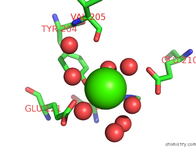
Mono view
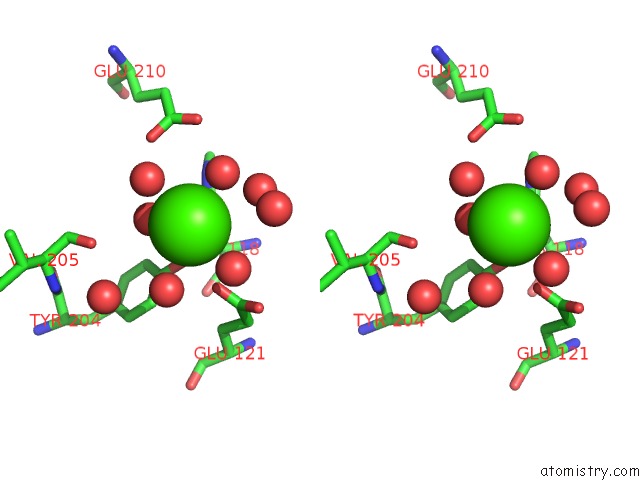
Stereo pair view
Mono view
Stereo pair view
A full contact list of Calcium with other atoms in the Ca binding
site number 2 of Crystal Structure of the Apo Form of Cmp-N-Acetylneuraminate Synthetase From Vibrio Cholerae within 5.0Å range:
|
Reference:
S.Bose,
D.Purkait,
D.Joseph,
V.Nayak,
R.Subramanian.
Structural and Functional Characterization of Cmp-N-Acetylneuraminate Synthetase From Vibrio Cholerae. Acta Crystallogr D Struct V. 75 564 2019BIOL.
ISSN: ISSN 2059-7983
PubMed: 31205019
DOI: 10.1107/S2059798319006831
Page generated: Tue Jul 16 09:23:17 2024
ISSN: ISSN 2059-7983
PubMed: 31205019
DOI: 10.1107/S2059798319006831
Last articles
Zn in 9J0NZn in 9J0O
Zn in 9J0P
Zn in 9FJX
Zn in 9EKB
Zn in 9C0F
Zn in 9CAH
Zn in 9CH0
Zn in 9CH3
Zn in 9CH1