Calcium »
PDB 6im3-6j43 »
6inz »
Calcium in PDB 6inz: Crystal Structure of Solute-Binding Protein Complexed with Unsaturated Hyaluronan Disaccharide
Protein crystallography data
The structure of Crystal Structure of Solute-Binding Protein Complexed with Unsaturated Hyaluronan Disaccharide, PDB code: 6inz
was solved by
S.Oiki,
B.Mikami,
K.Murata,
W.Hashimoto,
with X-Ray Crystallography technique. A brief refinement statistics is given in the table below:
| Resolution Low / High (Å) | 41.41 / 2.29 |
| Space group | P 21 21 21 |
| Cell size a, b, c (Å), α, β, γ (°) | 48.740, 97.501, 132.443, 90.00, 90.00, 90.00 |
| R / Rfree (%) | 16 / 21.7 |
Calcium Binding Sites:
The binding sites of Calcium atom in the Crystal Structure of Solute-Binding Protein Complexed with Unsaturated Hyaluronan Disaccharide
(pdb code 6inz). This binding sites where shown within
5.0 Angstroms radius around Calcium atom.
In total only one binding site of Calcium was determined in the Crystal Structure of Solute-Binding Protein Complexed with Unsaturated Hyaluronan Disaccharide, PDB code: 6inz:
In total only one binding site of Calcium was determined in the Crystal Structure of Solute-Binding Protein Complexed with Unsaturated Hyaluronan Disaccharide, PDB code: 6inz:
Calcium binding site 1 out of 1 in 6inz
Go back to
Calcium binding site 1 out
of 1 in the Crystal Structure of Solute-Binding Protein Complexed with Unsaturated Hyaluronan Disaccharide
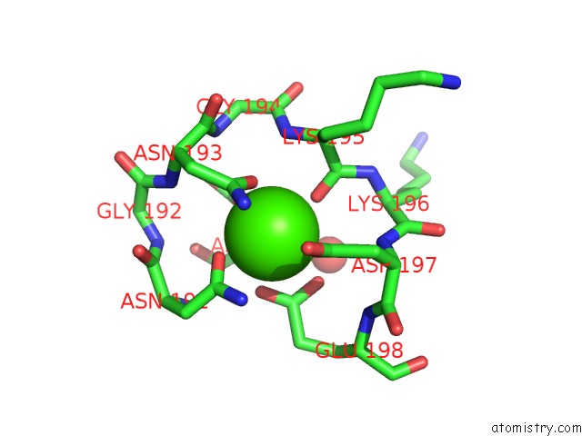
Mono view
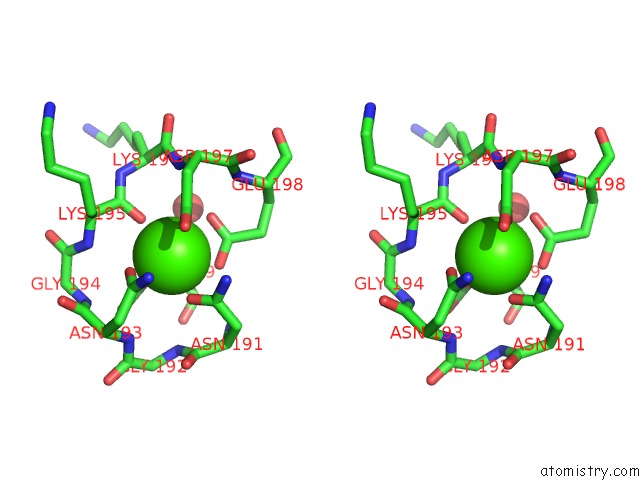
Stereo pair view
Mono view
Stereo pair view
A full contact list of Calcium with other atoms in the Ca binding
site number 1 of Crystal Structure of Solute-Binding Protein Complexed with Unsaturated Hyaluronan Disaccharide within 5.0Å range:
|
Reference:
S.Oiki,
M.Sato,
B.Mikami,
K.Murata,
W.Hashimoto.
Substrate Recognition By Bacterial Solute-Binding Protein Is Responsible For Import of Extracellular Hyaluronan and Chondroitin Sulfate From the Animal Host. Biosci.Biotechnol.Biochem. V. 83 1946 2019.
ISSN: ISSN 0916-8451
PubMed: 31204616
DOI: 10.1080/09168451.2019.1630250
Page generated: Tue Jul 16 09:31:09 2024
ISSN: ISSN 0916-8451
PubMed: 31204616
DOI: 10.1080/09168451.2019.1630250
Last articles
Zn in 9J0NZn in 9J0O
Zn in 9J0P
Zn in 9FJX
Zn in 9EKB
Zn in 9C0F
Zn in 9CAH
Zn in 9CH0
Zn in 9CH3
Zn in 9CH1