Calcium »
PDB 6im3-6j43 »
6isg »
Calcium in PDB 6isg: Structure of 9N-I Dna Polymerase Incorporation with Dg in the Active Site
Enzymatic activity of Structure of 9N-I Dna Polymerase Incorporation with Dg in the Active Site
All present enzymatic activity of Structure of 9N-I Dna Polymerase Incorporation with Dg in the Active Site:
2.7.7.7;
2.7.7.7;
Protein crystallography data
The structure of Structure of 9N-I Dna Polymerase Incorporation with Dg in the Active Site, PDB code: 6isg
was solved by
S.W.Linwu,
M.Maestre-Reyna,
M.D.Tsai,
Y.H.Tu,
W.H.Chang,
with X-Ray Crystallography technique. A brief refinement statistics is given in the table below:
| Resolution Low / High (Å) | 40.81 / 3.40 |
| Space group | I 2 3 |
| Cell size a, b, c (Å), α, β, γ (°) | 208.078, 208.078, 208.078, 90.00, 90.00, 90.00 |
| R / Rfree (%) | 25.2 / 30.2 |
Calcium Binding Sites:
The binding sites of Calcium atom in the Structure of 9N-I Dna Polymerase Incorporation with Dg in the Active Site
(pdb code 6isg). This binding sites where shown within
5.0 Angstroms radius around Calcium atom.
In total 5 binding sites of Calcium where determined in the Structure of 9N-I Dna Polymerase Incorporation with Dg in the Active Site, PDB code: 6isg:
Jump to Calcium binding site number: 1; 2; 3; 4; 5;
In total 5 binding sites of Calcium where determined in the Structure of 9N-I Dna Polymerase Incorporation with Dg in the Active Site, PDB code: 6isg:
Jump to Calcium binding site number: 1; 2; 3; 4; 5;
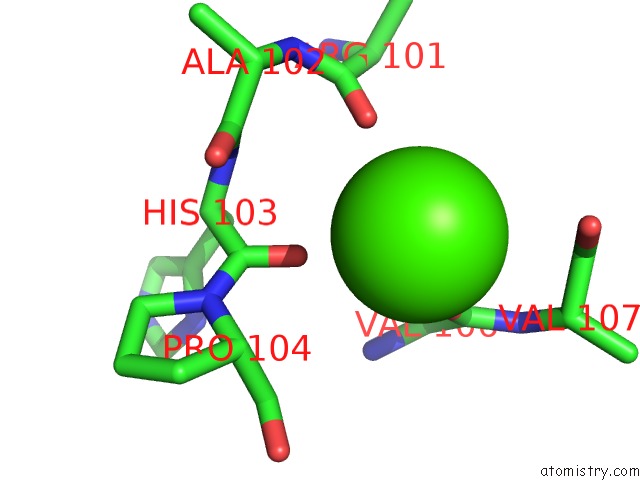
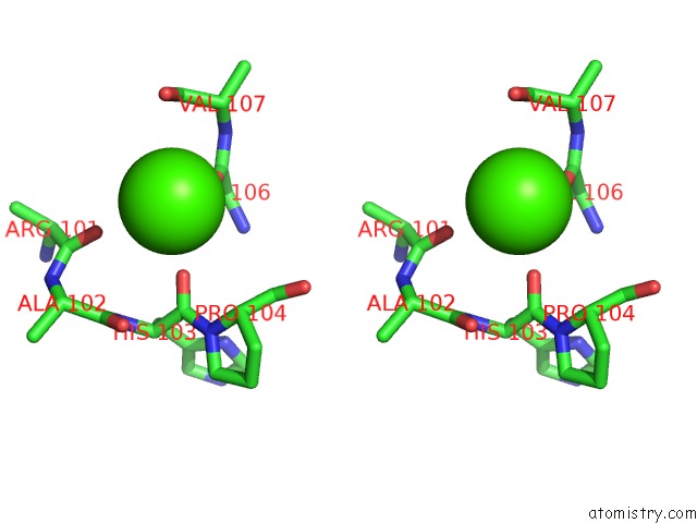
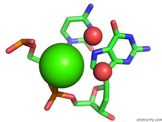
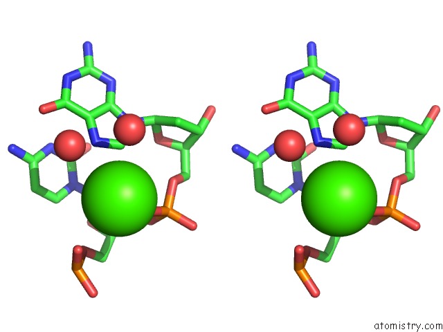
Calcium binding site 1 out of 5 in 6isg
Go back to
Calcium binding site 1 out
of 5 in the Structure of 9N-I Dna Polymerase Incorporation with Dg in the Active Site
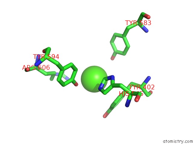
Mono view
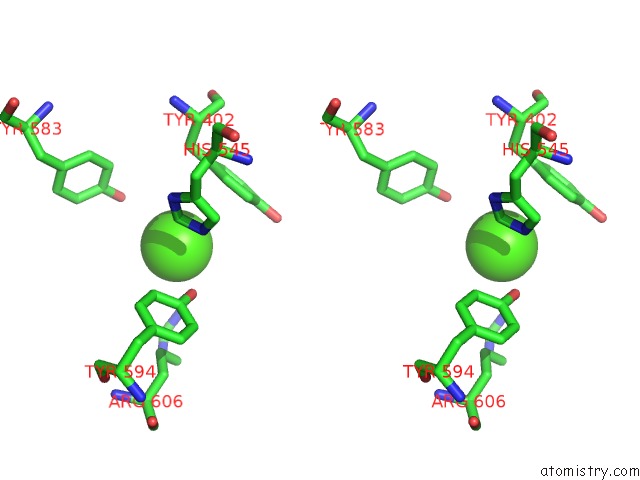
Stereo pair view
Mono view
Stereo pair view
A full contact list of Calcium with other atoms in the Ca binding
site number 1 of Structure of 9N-I Dna Polymerase Incorporation with Dg in the Active Site within 5.0Å range:
|
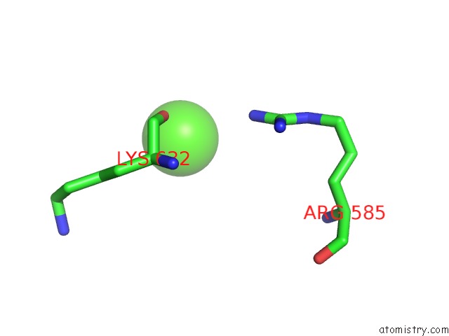
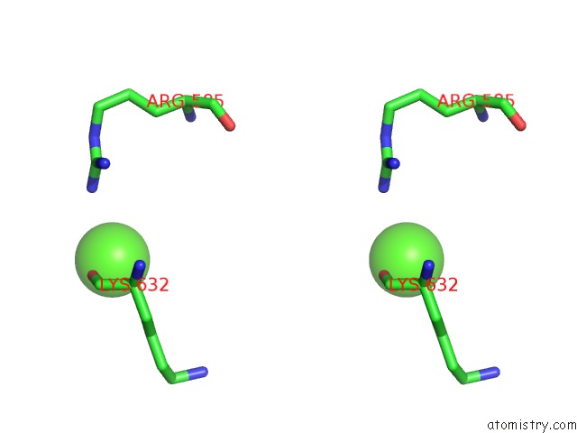
Calcium binding site 2 out of 5 in 6isg
Go back to
Calcium binding site 2 out
of 5 in the Structure of 9N-I Dna Polymerase Incorporation with Dg in the Active Site
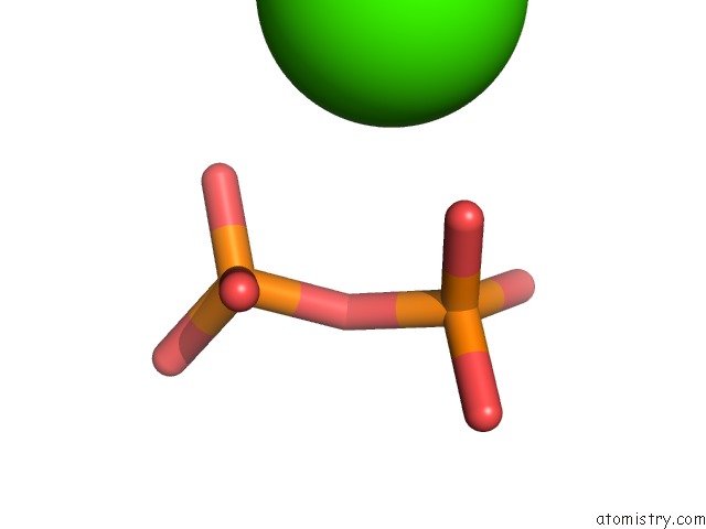
Mono view
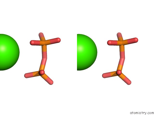
Stereo pair view
Mono view
Stereo pair view
A full contact list of Calcium with other atoms in the Ca binding
site number 2 of Structure of 9N-I Dna Polymerase Incorporation with Dg in the Active Site within 5.0Å range:
|
Calcium binding site 3 out of 5 in 6isg
Go back to
Calcium binding site 3 out
of 5 in the Structure of 9N-I Dna Polymerase Incorporation with Dg in the Active Site
Mono view
Stereo pair view
Mono view
Stereo pair view
A full contact list of Calcium with other atoms in the Ca binding
site number 3 of Structure of 9N-I Dna Polymerase Incorporation with Dg in the Active Site within 5.0Å range:
|
Calcium binding site 4 out of 5 in 6isg
Go back to
Calcium binding site 4 out
of 5 in the Structure of 9N-I Dna Polymerase Incorporation with Dg in the Active Site
Mono view
Stereo pair view
Mono view
Stereo pair view
A full contact list of Calcium with other atoms in the Ca binding
site number 4 of Structure of 9N-I Dna Polymerase Incorporation with Dg in the Active Site within 5.0Å range:
|
Calcium binding site 5 out of 5 in 6isg
Go back to
Calcium binding site 5 out
of 5 in the Structure of 9N-I Dna Polymerase Incorporation with Dg in the Active Site
Mono view
Stereo pair view
Mono view
Stereo pair view
A full contact list of Calcium with other atoms in the Ca binding
site number 5 of Structure of 9N-I Dna Polymerase Incorporation with Dg in the Active Site within 5.0Å range:
|
Reference:
S.W.Linwu,
Y.H.Tu,
T.Y.Tsai,
M.Maestre-Reyna,
M.S.Liu,
W.J.Wu,
J.Y.Huang,
H.W.Chi,
W.H.Chang,
C.F.Chiou,
A.H.Wang,
J.Lee,
M.D.Tsai.
Thermococcus Sp. 9°N Dna Polymerase Exhibits 3'-Esterase Activity That Can Be Harnessed For Dna Sequencing. Commun Biol V. 2 224 2019.
ISSN: ESSN 2399-3642
PubMed: 31240262
DOI: 10.1038/S42003-019-0458-7
Page generated: Tue Jul 16 09:34:10 2024
ISSN: ESSN 2399-3642
PubMed: 31240262
DOI: 10.1038/S42003-019-0458-7
Last articles
Zn in 9J0NZn in 9J0O
Zn in 9J0P
Zn in 9FJX
Zn in 9EKB
Zn in 9C0F
Zn in 9CAH
Zn in 9CH0
Zn in 9CH3
Zn in 9CH1